MRI/PET/electroencephalography (EEG) hybrid scanning has been achieved for the first time by a group of researchers from Jülich in Germany, according to a recent article published online by the Journal of Magnetic Resonance, which describes the work being done and makes a case for multimodal imaging at 3 tesla and 9.4 tesla.
The 3-tesla system is being used for basic neuroscientific research and clinical studies investigating the role of brain function in diabetes and obesity. The researchers have since begun to integrate EEG with a 9.4-tesla PET/MRI hybrid imager, the only such machine in existence.
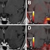
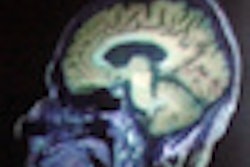
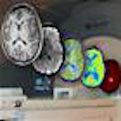
 The 3-tesla PET/MRI system at the Research Centre Jülich has been integrated with an EEG system to provide millisecond resolution of brain activity in tandem with molecular images obtained using PET, anatomy with MRI, and functional images based on the hemodynamic response with functional MRI. All images courtesy of Jon Shah, PhD.
Hybrid MRI/PET/EEG scanning adds temporal dimension to molecular, anatomical, and functional imaging. While PET is the gold standard for metabolic imaging and MRI provides high-contrast images of soft tissue, both provide largely static images of the brain. Furthermore, functional MRI (fMRI) provides only indirect evidence of brain activation or metabolism when an individual is presented with a task. The modality exploits the blood oxygenation level dependant (BOLD) effect that demonstrates hemodynamic responses to such tasks. "We're not measuring electrical activity with MRI; we're measuring a consequence thereof," explained lead author N. Jon Shah, PhD, a medical physicist at the Institute of Neuroscience and Medicine of the Research Center Jülich in Jülich, Germany (J Magn Reson, 10 December 2012).
EEG, on the other hand, can detect the activation of different structures in the brain with millisecond resolution. "We know the brain is not a static organ; it's doing things all the time. We wanted the temporal dimension and that's why we went to EEG," Shah said. The trimodal approach enables brain activation to be simultaneously observed with the hemodynamic and molecular information provided by MRI and PET, respectively.
Integrating off-the-shelf EEG
The trimodal scanner is based on a 3-tesla Tim Trio MRI system (Siemens Healthcare). A custom-built BrainPET insert enables simultaneous PET imaging. The insert contains 32 copper-shielded detector "cassettes" that use avalanche photodiodes, which are unaffected by magnetic fields, to detect the gamma photons emitted from the patient. While the insert was developed by Siemens, the researchers integrated the systems, developing accompanying software and debugging the hardware.
An off-the-shelf EEG system was then integrated with the PET/MRI system. This presented a number of technical challenges, on top of those associated with the integration of MRI with PET. For example, currents are induced in the electrodes attached to the patient by the switching gradient magnetic fields during a scan, which interfere with the ECG signal. Cardioballistic artifacts, where heart-related phenomena including the pulsation of blood vessels in the scalp, also degrade the quality of the EEG signal.
 Medical physicist N. Jon Shah, PhD, from the Institute of Neuroscience and Medicine (INM) at the Research Center Jülich.
However, both artifacts were successfully removed from sample EEG signals acquired in the scanner using methods including artifact average template subtraction. Photon scattering by the EEG electrodes and wires was found to have negligible attenuation effects on PET image quality.
The trimodal scanner is being used in three main ways by the researchers at the Jülich center. While their main focus is basic neuroscientific research, using the simultaneous scans to investigate brain function, the scanner is also producing data for the Imaging and Curing Environmental Metabolic Diseases (ICEMED) study that is investigating neurological aspects of diabetes and obesity. Clinically, the scanner is also in high demand for the preoperative assessment of brain tumor patients. The single procedure offered by the hybrid system has proved beneficial for the seriously ill patients who cannot tolerate more than one scan.
Trimodal imaging at 9.4 tesla
In related work, the researchers are also working toward an ultrahigh-field 9.4-tesla MRI/PET/EEG trimodal scanner. As a PET/MRI system alone and the only one of its kind, the scanner already provides superior submillimeter MRI resolution and 3-mm PET resolution of neurological structures such as the cerebellum. The researchers anticipate the extra information provided by the new technology will help identify potential biomarkers for the diagnosis and monitoring of conditions ranging from stroke to schizophrenia.
The 9.4-tesla system uses the same arrangement as the 3-tesla unity, with an MRI system accompanied by a BrainPET insert. The researchers are in the process of integrating the hybrid with an off-the-shelf EEG system. "Preliminary tests have already been carried out in the static field successfully," Shah said. Initial findings are under review for publication in NeuroImage.
© IOP Publishing Limited. Republished with permission from medicalphysicsweb, a community website covering fundamental research and emerging technologies in medical imaging and radiation therapy.