Navigators show promise for real-time organ tracking and breath-hold monitoring during MR-guided radiotherapy. They are a fast and precise method for monitoring and real-time tracking of anatomical landmarks by providing direct MRI feedback on anatomical changes for more precise radiation delivery.
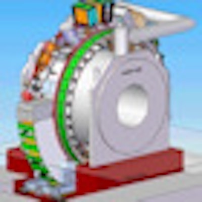
MRI offers unrivalled soft-tissue contrast for visualizing the target and surrounding organs during radiotherapy. Researchers at University Medical Center Utrecht in the Netherlands have built a prototype hybrid MRI-linac system. They are now investigating the use of continuous MR imaging during radiation delivery for gating or tumor tracking, which is described in an article published on 3 October by Physics in Medicine and Biology.
The MRI-linac, developed together with radiation oncology firm Elekta and Philips Healthcare, combines a 1.5-tesla MRI system and a 6-megavolt (MV) linear accelerator. To enable real-time motion detection during treatment, the researchers use pencil-beam navigators, fast one-dimensional MRI acquisitions that can be placed with arbitrary orientation in the anatomy. The team used in-house software to analyze the navigators, which are more commonly used for motion correction in diagnostic MRI of heart and lungs, for kidney tracking (Phys Med Biol, October 2012, Vol. 57:21, pp. 6797-6805).
 The MR-linac prototype. All images courtesy of Mette Stam and University Medical Center Utrecht.
"Pencil-beam navigators can be acquired in 15 milliseconds and therefore are suitable for very fast tumor motion detection," explained lead author Mette Stam, a medical physicist. "Furthermore, if a lower time resolution is good enough for motion detection, they can be interwoven with another MR sequence allowing, for example, simultaneous motion detection and functional imaging."
Motion tracking
The Utrecht team first evaluated the accuracy of navigator-based tracking using a water-fat phantom moving in a known one-dimensional periodic pattern. Standard pencil-beam navigators were acquired every 27 msec, with the center of the navigator positioned such that the air-water, water-fat, and fat-air boundaries were all tracked.
Results showed that the tracking consistency was within 0.5 mm. For an applied motion amplitude of 18.8 mm, for example, the measured amplitudes at the water-air, water-fat, and air-fat boundaries were 18.8, 18.7, and 18.7 mm, respectively. For an amplitude of 38.7 mm, the measured values were 38.9, 39.2, and 39.2 mm, respectively. The time required for navigator acquisition (15 msec) and image analysis (12 msec) is short enough to enable real-time radiotherapy guidance.
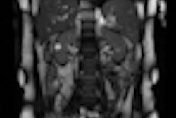
The researchers then tracked kidney motion in a volunteer, by acquiring alternating pencil-beam navigators (positioned in the direction of kidney motion) and 2D sagittal images of the kidney, during breath-hold and free breathing.
During the breath-hold, the mean deviation between the navigators and 2D images was 0.6 mm, with a maximum tracking deviation of 1.4 mm. The onset of breathing was seen in both the 2D images and navigators, though the amplitude differed between the two, likely due to kidney motion during the longer 2D acquisition (0.390 sec). Data recorded during free breathing also showed good overlap between breathing patterns detected by the two modalities.
 Overlap of 2D images and navigator measurements of the kidney during (a) breath-hold and the onset of breathing and (b) free breathing. During the breath-hold phase, both imaging modalities overlap and the phase of the free breathing is also equal.
Finally, the team assessed the use of navigators for breath-hold monitoring during a kidney treatment, examining seven patients with a renal lesion. A navigator was placed on the lesion in the direction of motion and patients performed an expiration breath-hold for as long as possible. Navigators were acquired every 25 msec until the patient restarted breathing.
The first stable navigator of the first breath-hold was used as a reference to analyze all navigators in five breath-holds of the same patient. If the result was within 2 mm of the reference position, the breath-hold was regarded stable. If not, the breath-hold was marked unstable and the treatment beam may be switched off.
As well as monitoring drift of the kidney during the breath-hold period, the navigators also detected the onset of breathing -- at which point the radiation can be switched off. The noise during tracking of kidney motion was larger than that seen in the phantom measurements, due to the fact that physiologic boundaries are less clear than phantom boundaries. The authors note that averaging the results of the last three navigators improved the accuracy.
 The five consecutive breath-holds of a patient set out after each other in one image. The green dots indicate the navigator results that are within 2 mm from the reference position and the red dots indicate the results further than 2 mm from the reference. If the edge is close to the threshold of 2 mm, the noise on the detection becomes apparent because the results seem to be oscillating around the threshold, shown as a mixture of red and green dots in the image.
Stam and co-authors conclude that this study demonstrates the suitability of navigators for rapid organ motion detection during breath-hold gated radiotherapy of kidney tumors, for example. They note that navigators are also suitable for target tracking during free breathing radiation delivery, particularly for reasonably rigid tumors that move along a straight line.
The researchers are currently working on the design of a future MRI-linac treatment for kidney tumors. "We are performing an observational MRI study on patients, examining the feasibility of both breath-hold and free-breathing gated radiation treatments," Stam told medicalphysicsweb.
© IOP Publishing Limited. Republished with permission from medicalphysicsweb, a community website covering fundamental research and emerging technologies in medical imaging and radiation therapy.