In Germany, a stroke occurs every three minutes. Eight out of ten strokes are ischemic, and in 30 to 40% of these cases, the cause cannot be established. Against this background, a German multicenter imaging study has analyzed the risks of instable carotid plaque.
Details about the study, called CAPIAS (Carotid Plaque Imaging in Acute Stroke), were presented at the German Radiological Society's annual meeting, the DRK.
The study aims to establish how often atherosclerosis is present in the internal carotid artery (ICA) of stroke patients. Preliminary data suggest approximately every third cryptogenic stroke may be caused by plaque breaking off and blocking vessels in the brain. Plaque may develop, and break off, in blood vessels either in situ in the brain, or it may be transported there from other body regions. The ICA plays a major role in this. A significant stenosis in the vessel can lead to a major stroke.
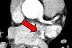
 Images of a patient aged 66, enrolled in the CAPIAS study in spring 2011 due to a right-hand infarction of the middle cerebral artery. The initial MRI showed a large and complex plaque with recent hemorrhage and lipid tissue (arrow), identified as complex type VI plaque based on categories of the American Heart Association. These plaques are considered particularly vulnerable, involving a high risk of cerebrovascular events. No stenosis is present. The lumen of the ICA is marked by the asterisk (*). All images courtesy of department of radiology, Ludwig Maximilian University Munich.
"This is a rather rare occurrence," explained Dr. Tobias Saam, from the Institute for Clinical Radiology of the Grosshadern Hospital at Munich University. "We assume a much more common phenomenon should be atherosclerotic plaques that hardly lead to a noticeable stenosis but are unstable, with pieces of plaque breaking off and traveling into the brain."
Exactly how often do unstable plaques occur in the ICA of stroke patients? This is the question being addressed by CAPIAS, which is being led by Saam in conjunction with Dr. Martin Dichgans from the Institute for Stroke and Dementia Research at Munich's Ludwig Maximilian University. Organizations participating in the investigator-initiated study include the Grosshadern Hospital and the Hospital Rechts der Isar in Munich, and soon Freiburg University Hospital will join them.
For CAPIAS, radiologists and neurologists work with a total of 300 stroke patients who show atherosclerotic changes of the ICA. Two questions are of key interest to Saam and his colleagues: to find out about the percentage of patients with unstable plaques in cryptogenic strokes, and to know whether unstable plaques coincide with a higher risk of recurrence.
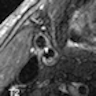
 Follow-up examination after 12 months shows a recent ulceration (point of arrow). The patient had a scotoma caused by a smaller thrombus. The most likely cause of this is plaque material that had broken off and led to the ulceration.
To find out, a high-resolution MRI examination of the carotid arteries is carried out within seven days of the stroke. After a year, this examination is repeated, and a brain MRI is performed. For black blood MRI, the signal from the blood flow is suppressed; the same sequences are in use for cardiac imaging. Dedicated surface coils are used; the so-called Blood Imaging Group in Munich, a team of graduate students, generates the necessary sequences for the study in the 3-tesla device. Imaging is done together with physicians and takes approximately 20-25 minutes for the carotids.
Based on this imaging method, type VI plaques can be identified. These plaques have a history of bleeding, clot formation, or rupture. One year after the launch of the study, data from the first 50 patients have now been evaluated to produce interim results.
In a third of cryptogenic stroke patients, type VI plaques were present in the carotid arteries. Out of the cases where neurologists had identified microvascular or cardioembolic causes of the stroke, only 10% showed these plaques. This may hint to the fact unstable plaques in the carotids are a potential cause of stroke which has hitherto been underestimated.
"We now have to wait for data to be confirmed further on. And we have to see what results look like a year further down," Saam said in Hamburg.
In case patients with unstable plaques do carry an increased risk of recurrence, interventional studies would need to be used to establish whether targeted early detection measures should be put in place. Likely therapies will be based on medication, or on invasive procedures to stabilize the plaques, he noted.