Use of extremely strong magnets in MRI can bring about high spatial resolution thus far only seen in light microscopes. Researchers at the University of Greifswald and Rostock in Germany demonstrated at the German Radiological Society's annual meeting, the DRK, that MRI microscopy provides as much information about the shape and growth of tumors as conventional histology.
MRI units of 7.1-tesla and more, used for research purposes and termed ultrahigh-field MRIs, can achieve resolutions of 40 micrometers and less. Using thin slice settings, radiologists can see images that used to be available only for tissue placed under the microscope. Cells and tiny blood vessels are visualized based on MR microscopy, with no biopsy required.
Dr. Paul-Christian Krüger and his team at the Institute for Diagnostic Radiology and Neuroradiology of the University Hospital in Greifswald have demonstrated the potential of the method. In a study conducted in collaboration with the ophthalmology clinic of the University Hospital of Rostock, they examined ten eyes resected because of malignant tumors of various types. They then compared results to those from histological analysis carried out at Rostock's pathology department.
 Section enlargement of a T-weighted sagittal image of a malignant uveal melanoma with infiltration of vitreous body, as well as extraocular growth infiltrating the sclera. All images courtesy of the radiology department, University Hospital Greifswald, and pathology department, University Hospital Rostock.
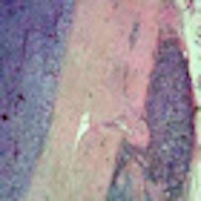
 Corresponding histological specimen (HE stain, enlargement 40x), correlating well with MR microscopy.
"MR microscopy visualizes the eye and its surrounding lipid tissue. This permits us to ascertain whether a tumor is growing into the surrounding tissue or not, which can play a decisive role in designing a therapy," he noted at the meeting. "Histological interpretation of the tumors produced, for all cases, identical results as in MR microscopy. For example, pathologists established that two tumors had grown into the surrounding tissue, congruent with our previous diagnosis based on MR microscopy."
To improve the quality and consistency of the new technology, histopathologic validation of imaging findings is required. Correlation between microMR images and histopathology can provide a better understanding of malignant eye processes and their treatment, he stated.
The next step is to establish the method in clinical routine, but work still needs to be done. The study was conducted with a 7.1-tesla device typically used in experimental research, but the group is now starting to do in-vivo exams in collaboration with a center with experience of using 7.1-tesla equipment in patients. A key step on the path toward clinical routine will be the development of dedicated surface coils that create an ideal magnetic field. Until now, coils are used for MR microscopy that have been designed for brain examinations. Surface coils can yield better images provided that movement of the head and eyes can be avoided. In addition, MR sequences will need to be shortened.
All these challenges can be resolved, Krüger said. As of this fall, in vivo studies will be launched in collaboration with the Max Delbrück Centre in Berlin. There, MRI physicists will create suitable coils that may enter the market at a later time.
In addition to early detection and precise diagnostics of tumors described above, applications include ocular lens implant positioning and cell proliferation for cataract surgery. Krüger proposed further application scenarios for the method: small joints, knee cartilage, and the inner ear are all visualized in a very detailed manner by MR microscopy.
"In general, [this method] is suitable for all structures that require high resolution rather close to the body surface," he concluded, raising the prospect of an end to biopsies at some stage.