Using certain pulse sequences, conventional 1.5-tesla MRI can visualize elbow ligaments that previously were not consistently visible in asymptomatic subjects, helping to diagnose patients with elbow instability, according to a study in the October issue of Radiology.
Swiss researchers noted that the additional information on the dimensions and signal intensity characteristics of ligaments and plicae in asymptomatic elbows may help avoid false-positive diagnoses in patients afflicted with elbow instability. The lead author of the study was Daniela Husarik, MD, from the department of radiology at University Hospital Zurich (Radiology, October 2010, Vol. 257:1, pp.185-194).
Though the assessment of ligaments is useful in evaluating patients with elbow instability, the authors cited previous research that found that some ligaments, such as the lateral ulnar collateral ligament, are not consistently visible on MRI or in dissected specimens of cadavers.
The prospective study enrolled 60 asymptomatic volunteers, which included 30 men and 30 women (median age, 32.8 years; range, 22-51).
MRIscans were performed on a 1.5-tesla system (Magnetom Symphony or Avanto, Siemens Healthcare, Erlangen, Germany) with patients in a prone position. A four-channel phased-array small extremity coil also was placed over the elbow joint.
The MR imaging protocol included five pulse sequences: transverse T1-weighted spin-echo, sagittal T2-weighted fast spin-echo, coronal fast spin-echo short-inversion-time inversion recovery (STIR), transverse intermediate-weighted with fat saturation, and coronal 3D water-excitation true fast imaging with steady-state precession (TrueFISP).
Three radiologists independently evaluated the MR images, which were presented to them in random fashion over an eight-week period. The trio analyzed the elbow ligaments -- which included the anterior ulnar collateral ligament, posterior ulnar collateral ligament, radial collateral ligament, lateral ulnar collateral ligament, and annular ligament -- for their respective degrees of visibility and signal intensity.
Visible ligaments
The 1.5-tesla MR images found the anterior ulnar collateral ligament and radial collateral ligament were completely visible in all 60 subjects (100%). The annular ligament was completely visible in 59 subjects (98%) and partially visible in one patient (2%).
In addition, the posterior ulnar collateral ligament was completely visible in 58 patients (97%) and only partially visible in two subjects (3%). The lateral ulnar collateral ligament was completely visible in 51 patients (85%) and partially visible in nine patients (15%).
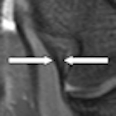
 |
| Coronal 3D MR image of an anterior ulnar collateral ligament (UCL) obtained with TrueFISP in a 27-year-old woman. The anterior UCL (arrows) has a striated signal intensity pattern. Images courtesy of Radiology. |
Increased signal intensity with fluid-sensitive sequences was seen in the anterior ulnar collateral ligament in nine (15%) of the 60 subjects and in the lateral ulnar collateral ligament in six patients (10%). Increased signal intensity was seen in the posterior ulnar collateral ligament in four subjects (7%), and it was found in the radial collateral ligament and annular ligament in one patient each (2%). The remaining ligaments had normal signal intensity on fluid-sensitive images.
 |
| Coronal STIR MR image of a 24-year-old man shows increased signal intensity in the proximal portion of the anterior UCL (arrows). |
MRIalso revealed the posterolateral plicae in 59 (98%) of the 60 subjects, while the posterior plicae was seen in 20 patients (33%).
Study limitations
A limitation of the study was the asymptomatic condition of the elbows analyzed, which may have affected qualitative assessments, according to Husarik and colleagues. "However, the facts that all ligaments could be measured and appropriate κ-values were obtained in the interobserver agreement test (0.48-1.00) indicate that the reading of the images was robust," they noted.
Even with this limitation, the authors concluded that elbow ligaments and the posterolateral plicae "can be seen on conventional MR images in most asymptomatic subjects. Most asymptomatic ligaments are thinner than 4 mm, and most plicae are thinner than 3 mm."
The clinical benefit from the results is that the study provides "quantitative values that may be useful for evaluating symptomatic elbows," they wrote.
By Wayne Forrest
AuntMinnie.com staff writer
September 27, 2010
Related Reading
JOCD: Imaging growth plate injury means scrutinizing bone-joint stability, September 4, 2007
Part I: Choosing between MR and US in musculoskeletal imaging, September 7, 2006
US-guided needle tenotomy benefits tennis elbow, January 24, 2006
Radial head-capitellum view best as adjunct in elbow x-ray, March 9, 2005
The bends and flexures of forearm and elbow x-ray positioning, November 21, 2002
Copyright © 2010 AuntMinnie.com