Ultrahigh dose rate ionizing radiation delivered in submillisecond pulses causes less damage to healthy tissue, with comparable tumor control to that seen from the continuous irradiation used in clinical radiotherapy. That's the conclusion of a preliminary study by researchers in France (Science Translational Medicine, 16 July 2014, Vol. 6:245, pp. 245ra93).
The findings have potential implications for clinical treatment approaches already using high dose rates and short irradiation times, such as scanned proton beams that deliver intensity-modulated proton therapy (IMPT). In the long term, they may also pave the way for pulsed photon treatments with improved therapeutic ratios.
.png?auto=format%2Ccompress&dpr=2&fit=max&q=70&w=700) A group photo of the Institut Curie -- INSERM U612 Genotoxicology, Signalisation, and Experimental Radiotherapy unit. Vincent Favaudon is in the center at the front.
A group photo of the Institut Curie -- INSERM U612 Genotoxicology, Signalisation, and Experimental Radiotherapy unit. Vincent Favaudon is in the center at the front."I'm confident that the process will find applications in the clinics either to reduce the level of complications, or increase the dose for a higher antitumor efficiency, or both," said Vincent Favaudon, first author and radiobiologist in the Inserm Unit 612 at the Institut Curie in Orsay, south of Paris.
Favaudon and his colleagues carried out the research following an in vitro study they undertook at the turn of the century. Mammalian cell cultures showed differences in radiation susceptibility, depending on whether they were exposed to pulsed or continuous radiation, and the findings were supported by subsequent cytogenetics studies by other groups.
In the meantime, treatment delivery techniques have been evolving. Flattening filter-free photon beams have dose rates several times that of conventional megavoltage photon beams. Accelerators delivering scanned proton pencil beams with dose rates around three orders of magnitude greater than conventional photon beams in short time intervals are also becoming a reality.
For example, due online in 2015, the pencil-beam facility at the Institut Curie's Proton Therapy Centre (also in Orsay) will deliver on the order of 70 Gy/s with irradiation times per beam spot of around 10 ms. Such developments motivated Favaudon and his colleagues to investigate further.
Comparing tissue response
The researchers compared the difference in radiation response between mice irradiated with "FLASH" and "CONV" regimes. FLASH used a 4.5 MeV electron beam with pulses less than 500 ms at an ultrahigh dose rate of more than 40 Gy/s, delivered using a preclinical research accelerator. CONV involved continuous irradiation at a dose rate less than 0.03 Gy/s, using either the same linac used to deliver FLASH, 662 eV gamma photons from a caesium-137 source, or 200 kV X-rays. The three sources were used for logistical reasons, with corrections made for the differing relative biological effectiveness of their beams.
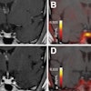
Responses in normal lung tissue and tumor were investigated using a C57BL/6J mouse model. Lungs were chosen over other anatomy for the parallel with lung cancer patients, where lung fibrosis is one late side effect of radiotherapy. Additionally, lung fibrosis in the model is the most extensively documented radiation-induced complication in laboratory animals.
When 240 healthy mice were divided into FLASH, CONV, and control groups, greater normal lung tissue sparing was observed in mice receiving FLASH, with no signs of lung fibrosis after 20 Gy. Those who received CONV had fibrosis after receiving 15 Gy. Additionally, histological testing of the irradiated lung tissue revealed 30 Gy delivered with FLASH versus 7.5 Gy delivered with CONV produced the same extent of apoptosis in smooth muscle and epithelial cells.
When tumor growth following irradiation was observed -- in subcutaneously implanted human breast and head-and-neck tumors as well as in in situ mouse lung tumors -- FLASH and CONV performed comparably.
Further work needed
Clinical trials remain some way off with no commercially available clinical linacs capable of delivering pulsed ultrahigh dose rate electron or photon beams. "Overcoming this limitation is not a short hop, but proton pencil-beam scanning facilities might offer an alternative," Favaudon said, adding that his group plans to use the Institut Curie's new proton pencil-beam facility in their ongoing research.
Further radiobiological studies are also needed to identify the mechanism by which FLASH spares healthy tissue. The group are investigating this with comparisons of different organs and studies at molecular, subcellular, and cellular levels and expect to publish more results within the next six months. They are open to interdisciplinary collaborations with other institutions.
© IOP Publishing Limited. Republished with permission from medicalphysicsweb, a community website covering fundamental research and emerging technologies in medical imaging and radiation therapy.