In a step toward the clinical use of ultrasound-mediated doxorubicin (DOX) delivery, a joint French-Dutch research project has identified the ultrasound characteristics required to release the chemotherapy drug from temperature-sensitive liposomes. The team also verified that the acoustic technique does not alter the drug's properties.
Targeted delivery of chemotherapy using nanoparticles is a promising and potentially powerful technique for treatment of cancer. Passive approaches exploit the selective accumulation of nanoparticles in the tumor due to its abnormal vasculature, but this has several limitations, including the fact that such accumulation doesn't occur in all tumors. Active delivery approaches are more promising and include the use of mild hyperthermia to release the drugs once in situ from temperature-sensitive nanoparticles.
Focused ultrasound (FUS) offers a targeted and noninvasive way to do this. Lead author Jean-Michel Escoffre, PhD, and co-workers -- from the Imaging and Brain INSERM unit at the Université François Rabelais de Tours in France and Eindhoven University of Technology in the Netherlands -- investigated the FUS parameters required to release DOX and its properties after release. They used a water bath heated to 37°C to mimic in vivo conditions. A small glycerol-filled vessel positioned in the bath held a sample holder containing the DOX solution positioned at the focus of a 1 MHz single element transducer (Physics in Medicine and Biology, 2013 November 21, Vol:58[22], pp. 8135-8151).
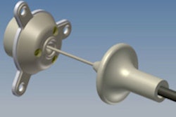
 The researchers induced hyperthermia in DOX in two ways. The first used FUS to heat the glycerol reservoir surrounding the sample holder, which in turn heated the holder and its contents. The second heated the water bath by conventional means, in turn heating the glycerol reservoir and the sample holder. All images courtesy of Jean-Michel Escoffre, PhD.
The right acoustic parameters
In a first experiment, the researchers established the acoustic parameters needed to heat a given volume in the sample holder to 42°C or above -- the phase transition temperature of the liposomes -- in order to release the encapsulated DOX. Results showed that a pulse repetition period of 1 ms, a pulse length of 400 cycles, and a peak negative pressure of 2 MPa over 10 minutes was sufficient to raise 115 mm3 of the solution above the threshold temperature.
A second experiment investigated the variation in DOX release from the liposomes with peak negative pressure and hyperthermia duration, measured by detecting the fluorescent intensity of the DOX upon release. When compared with DOX released by conventional heating of the surrounding water bath, the two methods proved equivalent: the same amount of DOX was released by each. DOX release only occurred for FUS with a peak negative pressure greater than approximately 1.5 MPa.
In a comparison with unencapsulated DOX, three further experiments investigated the uptake of liposome-encapsulated DOX by glioblastoma tumor cells and, after treatment, the viability of the cells and the growth rates of 3D cell cultures (known as multicellular tumor spheroids). In each experiment, again it made no difference whether the encapsulated DOX was heated directly with FUS or using the water bath.
','dvPres', 'clsTopBtn', 'true' );" >
A temperature rise from 37°C to 42°C showed a statistically significant increase in the uptake of encapsulated DOX by the cells and a corresponding decrease in cell viability and increase in spheroid doubling time. For example, the spheroid doubling time was more than two times longer at 42°C compared with 37°C. Meanwhile, when the drug was in its free form, the same temperature change resulted in no significant change in DOX uptake, cell viability, or spheroid growth.
The researchers conclude that in vitro at least, FUS is as efficient as conventional water bath heating in the release of encapsulated DOX and that the ultrasound does not change its pharmacological behavior.
The mechanism of drug release is still unknown, however. Potential mechanisms include the formation of heat-induced nanopores and the rupture of the liposomes walls by inertial cavitation. In ongoing work, the group aims to uncover the details of the drug release mechanism, as well as perform in vivo studies to assess the efficacy and safety of the approach for cancer treatment.
"The study of mechanisms involved in the drug release from the nanoparticles is a key element to improve the efficacy of ultrasound-triggered drug delivery, as well as the therapeutic efficacy and the safety of this particular procedure," Escoffre said, who now works at the University Medical Center Utrecht in the Netherlands.
© IOP Publishing Limited. Republished with permission from medicalphysicsweb, a community website covering fundamental research and emerging technologies in medical imaging and radiation therapy.