Hadron therapy is an advanced cancer treatment that employs beams of protons or carbon ions to deliver dose with submillimeter accuracy. The extreme precision of this approach, however, means that treatment can be compromised if the range of the charged particles differs from the treatment plan. As such, range verification techniques are under investigation, with PET imaging of positron emitters generated by the treatment beam already in clinical use.
The Innovative Solutions for Dosimetry in Hadrontherapy (INSIDE) collaboration is developing a bimodal imaging system for verifying head-and-neck treatments. The system combines an in-beam PET scanner with an integrated charged particle tracking system. The in-beam PET part of the system is now being clinically validated at the Centro Nazionale di Adroterapia Oncologica in Italy, and was tested during a patient treatment for the first time on 1 December 2016 (Journal of Medical Imaging, 2 December 2016, Vol. 4:1. pp. 011005).
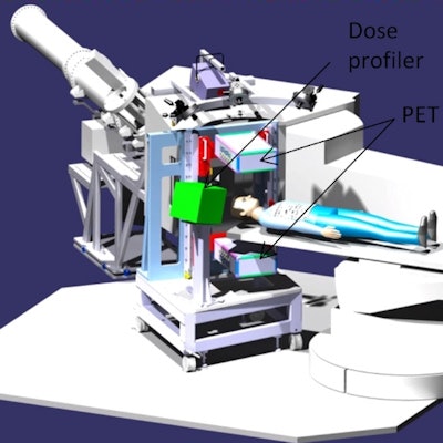
 A schematic showing the complete INSIDE bi-modal system.
The INSIDE research project was launched in 2013 under the PRIN (Relevant National Interest Projects) program of the Italian Ministry of Education, University and Research (MUIR). INSIDE is coordinated by the University of Pisa, in collaboration with the Universities of Turin and La Sapienza of Rome, Bari Polytechnic University, and INFN.
The bimodal INSIDE imaging system comprises an in-beam PET scanner made of two opposing planar heads and a dose profiler for charged particle tracking. In-beam PET scanners that operate during delivery of the beam have higher sensitivity and measurement capabilities than in-room or offline PET systems. The PET detector is based on solid-state photodetectors (silicon photomultipliers) with high granularity and one-to-one coupling to lutetium fine silicate (LFS) pixelated scintillating crystals. Custom software performs the system calibrations, monitors online performance and reconstructs the PET images.
A single events online monitoring functionality is used to separate in-spill and inter-spill data, by measuring the event rate and comparing it with a threshold. Real-time reconstruction capability provides on-the-fly imaging of the activity map during beam delivery. This enables almost real-time range assessment, with the possibility of providing feedback to the beam delivery system, such as an emergency beam stop.
System testing
Principal investigator Maria Giuseppina Bisogni, of the University of Pisa's Department of Physics, and colleagues, tested and commissioned the system. They conducted preliminary tests with different irradiation settings using polymethyl methacrylate (PMMA) phantoms (solid phantoms and ones with small air and bone inserts).
 The INSIDE in-beam PET system installed in the treatment room at CNAO in Pavia, Italy.
The aim of these tests was to perform a first evaluation of system performance by separately analyzing datasets acquired during in-spill and inter-spill irradiation phases. The team also used the measurements to test the simulations under easily reproducible conditions. Particle range was measured in terms of activity range induced in both homogeneous and heterogeneous phantoms from primary beams of fixed and variable energies.
For the first set of measurements on PMMA phantoms, the in-spill image was noisier than the inter-spill image, due to the radiation-induced background. The authors reported that the proximal rise and distal fall-off edges for inter- and in-spill data were in good agreement (to within less than 1 mm for the distal fall-off), and that reliable activity range measurements could be performed during both the inter-spill and in-spill periods.
"Increasing the spill time and accordingly shortening the inter-spill time shortens the duration of the treatment, which is a benefit to the patient. However, the in-spill time will be dominant compared with the inter-spill time," Bisogni told medicalphysicsweb. "Initial data showed that the quality of the in-spill PET images was worse than that of the inter-spill images, with the noise due to random coincidences much higher than in the inter-spill images. The increase of random coincidences in the in-spill images is due to the background radiation that is promptly emitted during the interaction of the beam with matter."
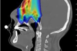
 PET images obtained when irradiating a PMMA phantom (10 x 10 x 20 cm, placed at the isocenter, the long side is parallel to the beam direction with the beam entering from the top) with a proton beam of 144 MeV (total dose 4.5 Gy). The left image was obtained during the inter-spill phase, the right image during the in-spill phase.
"Unfortunately, standard methods for random correction cannot be applied in this case," she added. "They require that the event rate is constant in time, while the background events (prompt gammas and neutrons) follow the time structure of the accelerated beam, a periodic structure in which the protons are accelerated and extracted in bunches of about 140 ns width at a frequency of 1.45 MHz. In this respect, our research is now focused on conceiving and developing smart techniques for suppression of the accelerator-induced random events to improve the signal-to-noise ratio of the in-spill PET images."
To assess system functionality under clinical conditions, the team also performed measurements on an anthropomorphic phantom. They created an image of the CT fused with the reconstructed activation map generated in the phantom by a proton beam shaped according to a real treatment plan, with proton energies ranging from 74 MeV to 134 MeV. The PET image was acquired and reconstructed within a 185 second irradiation session in a setup reproducing a clinical treatment -- making this test a milestone for the INSIDE project. The team is now conducting simulations of real patient cases.
Clinical validation of the INSIDE prototype began in December, with the researchers monitoring the treatment of a patient with cancer of the lacrimal glands. "The test was successful," Bisogni said. "We repeated it the next day with comparable results." The statistical uncertainties of INSIDE will be assessed as clinical validation continues. Other future plans include developing a motion-controlled positioning system that would overcome a limitation of the existing manual positioning system.
© IOP Publishing Limited. Republished with permission from medicalphysicsweb, a community website covering fundamental research and emerging technologies in medical imaging and radiation therapy.