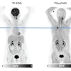
PET/CT with FDG-labeled leukocytes may detect infection in patients with acute pancreatitis, according to a study published in the August issue of the Journal of Nuclear Medicine.
The study from the Postgraduate Institute of Medical Education and Research in Chandigarh, India, included 41 patients (28 men and 13 women, ages 21-69) with acute pancreatitis and radiological evidence of fluid collection in or around the pancreas (JNM, August 2014, Vol. 55:8, pp. 1267-1272).
Leukocytes (white blood cells) were separated from the patient's venous blood, labeled with FDG, and reinjected intravenously. PET/CT imaging was performed two hours later. A final diagnosis of infection was based on microbiological culture of fluid aspirated from the collection. Patients were managed with supportive care and antibiotics, and percutaneous drainage or laparotomy was performed when indicated.
Increased FDG uptake was seen in the collection in 12 (29%) of the 41 patients; 10 had culture-proven infection and underwent percutaneous drainage, and aspiration was unsuccessful in two patients, the researchers found. PET/CT was negative for infection in 29 patients (71%). Of the 29 patients, 25 had negative fluid cultures, and aspiration was unsuccessful in four. Sensitivity, specificity, and accuracy of the scan were 100% in the 35 patients for whom fluid culture reports were available.
By using this new technique to diagnose infection in patients with pancreatic fluid collection, individuals are spared empiric antibiotic therapy or radiological intervention followed by time-consuming microbiological workup, lead author Dr. Anish Bhattacharya said in a statement.
The results also suggest that PET/CT with FDG-labeled leukocytes could be useful for detecting or ruling out active infection in any part of the body, Bhattacharya added.


