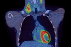
How can we estimate the risk of inducing second cancers in patients receiving radiotherapy? Ionizing radiation is known to cause cancer, and the risk of inducing second cancers in patients undergoing radiotherapy cannot be ignored.
Speaking at the Clatterbridge Course on Radiobiology and Radiobiological Modelling in Radiotherapy, held last month in Port Sunlight, U.K., Geoff Lawrence, PhD, head of imaging physics from Clatterbridge Cancer Centre in Wirral, U.K., discussed how the effects of irradiation can best be modeled and predicted.
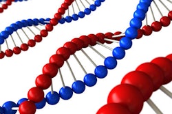
Lawrence began by describing the mechanisms of radiation carcinogenesis. Radiation acts as an initiator, he explained, causing DNA damage that, if it doesn't kill the cell and isn't repaired, results in a population of mutated cells. Following a multistage process, these abnormal cells may eventually give rise to clinical cancer.
So, is it possible to estimate the risk of second cancer induction due to radiation? Epidemiological studies can shed light on potential risk levels, with the largest source of information arising from the Life Span Study of Japanese atomic bomb survivors. This study, with nearly 70 years follow-up, clearly demonstrates excess cancer risk at organ doses above 50-100 mSv, with leukemia cases appearing within a few years of radiation exposure and solid tumors dominating in the long term.
The excess relative risk -- defined as a percentage increase in the underlying risk of developing cancer -- was higher for those exposed when young. "If irradiated at a young age, the lifetime risk is much higher than for those irradiated when older, and the risk is higher for females," Lawrence noted.
Treatment specific
While the Japanese Life Span Study (LSS) provides the greatest amount of data, it may be possible to gain more focused information by examining cancer incidence following radiotherapy. Lawrence cited a study of second cancer induction after radiotherapy for breast cancer. Results showed a lower risk than expected from the study data, demonstrating the impact of fractionation.
Dose level can also affect the outcome. At low doses and dose rates, risk increases linearly or linear-quadratically with dose. At higher doses, however, another effect comes into play: more cells are killed by the radiation and the gradient of the dose-risk curve is reduced. "The risk coefficients from atomic bomb survivors' study and other low-dose epidemiological studies may be taken as an upper bound to the risk coefficients for radiotherapy," Lawrence told the delegates.
Many studies have revealed an increased incidence of second primary cancers after radiotherapy. These include large, statistically powerful cohort studies, as well as case-controlled studies that match patients who develop second cancers against those who do not. Lawrence notes that risk estimates derived for the general population must be used with care, as cancer patients may have different underlying risk factors: They tend to be older and may have significant genetic, environmental, or lifestyle differences.
He described an example study of patients with Hodgkin's disease, an important group as such patients are young when diagnosed, are usually cured, and have a long life-expectancy. A cohort of 1,319 patients was treated using radiotherapy, with or without chemotherapy. Of these, 181 developed second malignancies, including 23 acute leukemias and 131 solid tumors.
In this study, excess risk continued to rise even after 15 to 20 years of follow-up. "We see risk increase and show no sign of flattening out -- this is a problem and it needs to be taken seriously," Lawrence said. He noted that the relative risk was lower for radiation therapy alone than for combined treatment, and the risk increased with radiation field size.
Looking more specifically at the induction of breast cancer after radiotherapy for Hodgkin's disease, risk was seen to increase with the radiation dose to the site where the tumor developed. "For patients treated with radiotherapy alone, the relative risk increased with dose, even to the highest dose levels," Lawrence said. Interestingly, chemoradiotherapy patients had greatly reduced risk compared with radiotherapy alone, due to protective hormonal changes associated with early menopause, thus emphasizing the need to understand all factors that may affect outcome.
Another confounding issue, Lawrence explained, is that findings are often based on historical data from treatments performed 10 to 20 years ago, or more. Since then, radiotherapy technologies have evolved greatly and the challenge now is how to use this historical information to predict risk from today's treatments. He showed data from a recent study of prostate cancer patients that suggest the risks of second cancer from some modern radiotherapy techniques may be lower than expected from older treatments.
Risk models
Based on the results of these epidemiological studies, and the many others that have been performed, is it possible to determine the risk of second primary cancers after radiotherapy? Lawrence noted that while the LSS provides an abundance of data regarding exposures of between 50 mSv and 2.5 Sv, less is known about the form of the risk function for lower and higher doses.
There's also real need for risk models to account for different types of exposure -- such as various dose levels and rates, fractionation, and heterogeneous dose distributions. Lawrence introduced the idea of the organ equivalent dose (OED), the uniform dose with the same risk of radiation-induced cancer as a delivered inhomogeneous dose distribution. If OED is a surrogate for risk, then it should be possible to compare risks from different techniques.
Lawrence presented details of several studies that aim to determine relationships between risk and dose parameters. One study proposed a function that rises roughly linearly at lower doses and at some point turns down due to killing of mutated cells. Another proposed model predicted that cell killing would reduce the risk of second cancer at higher doses, but also that fractionation provides an increased ability for repair of sublethal damage to mutated cells, thereby increasing the risk at high doses.
Collating all information, Lawrence suggested that for low-dose regions distant from the treatment volume, "We should probably go with the linear-no-threshold hypothesis, and apply organ-specific risk coefficients." In high-dose regions close to the target, meanwhile, "We need a more sophisticated nonlinear model, with parameters fitted from epidemiological evidence if available."
"We still need to establish the best way to establish risks of second cancer from radiotherapy," he concluded. "We've got some promising models, but we will only know if they are right in 10 to 20 years' time. So we need to use what we've got intelligently, but also to keep on recording outcome data to see if our models are reliable."
In discussion after his presentation, Lawrence agreed that the risks of second cancers must be kept in perspective and considered in the context of the usually great benefit that the patient is expected to get from radiotherapy, which may be life-saving.
© IOP Publishing Limited. Republished with permission from medicalphysicsweb, a community website covering fundamental research and emerging technologies in medical imaging and radiation therapy.