Visualization experts and radiologists from Norrköping and Linköping, Sweden, have contributed their renowned expertise and skills to a pioneering project designed to shed new light on Egyptian mummies and improve education of museum visitors.
Staff from the Museum of Mediterranean and Near Eastern Antiquities (Medelhavsmuseet) in Stockholm is working with a group of researchers from the Interactive Institute Swedish ICT to digitally scan human mummies as part of the preparations for a permanent exhibition.
The results for one of the mummies, the Egyptian priest Neswaiu, are now on display in the form of a digital visualization table in a room beside the real mummified remains and coffins. Using the table, visitors can virtually open the two coffins and then unravel each layer of the mummy from his highly decorated cartonnage, or outer layer, right down to the skeleton. They can also cut a cross-section through the multiple layers of the coffins and body.
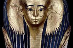
 The mummy of Neswaiu, who lived in the third century B.C. at the temple of the god Montu in Thebes (modern-day Luxor), was captured in 3D using a combination of dual-energy CT scanning, photogrammetry, and laser scanning. His remains were donated to the Medelhavsmuseet in 1928, when it first opened. Image courtesy of Interactive Institute Swedish ICT.
"We have been working with our partner Anders Persson and his fantastic team at the Center for Medical Image Science and Visualization in Linköping, Sweden," said Thomas Rydell, studio director of Interactive Institute Swedish ICT in Norrköping. "The software used is an in-house visualization engine called Inside Explorer, which you can read about here: www.tii.se/projects/insideexplorer. In conjunction to the CT scanning, we also used laser scanning and photogrammetry."
To examine the mummies, Dr. Anders Persson, PhD, and colleagues use a dual-energy CT scanner (Somatom Definition Flash, Siemens Healthcare). In the box below, you can see his practical tips for anybody who wishes to study Egyptian mummies.
Initially, Inside Explorer was developed for use in hospitals and by medical students, but Rydell and his colleagues have gone on to work with the British Museum on a visualization table for the museum's Gebelein Man, a 5,500-year-old mummified Egyptian man. They have also worked with London's Natural History Museum, Chicago's Field Museum of Natural History, and the Smithsonian in Washington, DC.
The Neswaiu project represents the team's most advanced work yet, combining data from both CT scanning and photogrammetry, by which 2D pictures of the coffins and mummy were taken from multiple angles to build a 3D surface model using the Autodesk Recap Photo software, according to Rydell. CT scanning provides information about the interior of the mummy, but does not offer any color or surface information, and laser scanning and photogrammetry give details about the surface, textures, and colors.
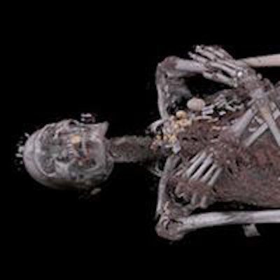
 Visitors to Stockholm's Medelhavsmuseet can now digitally unwrap the mummy of an Egyptian priest. Data from the CT and 3D surface scanning have been combined in Inside Explorer -- an interactive visualization touch table developed by Interactive Institute Swedish ICT. Inside Explorer creates a photorealistic digital representation of the mummy that allows the visitors to explore all the layers of the mummy inside and out -- from the small carvings on the sarcophagus to the anatomy -- as well as the artifacts wrapped together with the body. Image courtesy of Interactive Institute Swedish ICT.
The digital autopsy has added new details about the mummy. He might have lived until he was 60 years old, which was relatively old in ancient Egypt, and he could have died from an infection in one of his teeth, which affected the bone and may have caused blood poisoning.
Furthermore, the process has given insight into the mummification process; the cut where the internal organs were taken out is visible, and it's possible to see the wrapped packages of intestines, lungs, and the liver put back inside the body. Also, one can see how the cut was resealed and an amulet in the shape of the embalmer's two fingers was put across the cut to protect it. Scanning also showed in 3D where 120 amulets found on his body were placed, including a falcon-shaped one. The data were used to make a 3D print of a mould and then cast an exact replica of the falcon while leaving the original undisturbed on the mummy.
 A mummy from Medelhavsmuseet in Stockholm is scanned by a dual-energy CT scanner at the Center for Medical Image Science and Visualization. Image courtesy of Karl Zetterström, Världskulturmuseerna.
This 3D digitization project has been part of Projektarena IVM and is in part financed by the European Regional Development Fund. To watch a short video about it on the BBC website, click here.