The two key technological advances of CT are aimed at enhancing the contrast of the image to reduce the quantity of contrast product injected, and reducing radiation dose. The reduction in acquisition time generates fewer artifacts, and the increase in the number of detectors opens up another means of acquisition. There are three principle axes of development applicable to urinary tract investigation.
Spectral energy (dual energy)
The benefit of dual energy acquisition with a polychromatic beam (e.g., 80 kVp and 140 kVp) consists in allowing distinction of two elements that have similar or close densities in classic acquisition (mono-energy: 120kV). This is due to their specific attenuation profiles.
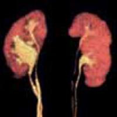
Acquisition in spectral energy provides further tissue characterization compared with mono-energy. The "monochrome" image (Fig. 1) is a hybrid image that associates intense contrast due to low kV image and low noise due to high kV image. Different reconstructions of the same slices obtained from a single spiral acquisition are based on the attenuation coefficient for each component (iodine, calcium, water, uric acid, fat, etc.), which generates a specific image for each of them.
 Monochrome image at 50 kV, coronal slice, arterial phase, contrast product used: 270 mgI/ml-50cc or 13.5 gr of iodine. All images courtesy of JFR.
This type of acquisition comprises extra tissue characterization adaptable to each clinical context through the potential information provided: noninjected image (water image), iodine image, calcium image, or that of uric acid or even fat.
Radiation dose is a little higher than in classic acquisition, but this means the phase without injection, provided by the water image, can be avoided, thus limiting the number of spirals. Recently, the use of iterative reconstruction techniques has reduced radiation.
In urinary pathology, several clinical applications are emerging: detecting or ruling out contrast take-up in a hemorrhagic zone; achieving an optimal parenchymal contrast and establishing internal vascular architecture of a tumor; improving image contrast; and finally, making the differential diagnosis between uric acid, calcium, or mixed stones (Fig. 2).
 Sequential image acquisition (five volumes) of a 62-year-old woman. Body mass index: 33.8 -- uro CT: bilateral pyeloureteral duplicity with lithiasis within the lumbar ureter of the lower right pyelon with overlying spasms. Volume rendering and maximum intensity projection reconstruction -- continuity of the different segments of the ureter are satisfactory -- dose length product 149 mGy.cm.
The impact of x-rays on iodine, factoring in its high atomic number, is principally due to a photoelectrical effect. For this reason, the attenuation of structures containing iodine is higher at 80 kV (around 100% higher), than at 140 kV. Therefore, with lower kV it is possible to reduce the quantity of iodine injected by more than 60% while retaining an excellent image quality in terms of noise signal and contrast with diagnostic information, particularly at 50 kV (Fig. 1).
Therefore, this type of acquisition in routine clinical practice is advantageous for the patient with moderate renal failure, while maintaining hyper hydration in a patient without cardiac failure, and obviating the taking of potentially nephrotoxic medicine.
This acquisition helps to better evaluate the degree of renal arterial stenosis by ruling out calcium plaque through iodine imaging. Furthermore, it provides quantification of iodine in µg/cm3 or the quantity of water in mg/cm3 in the renal parenchyma, with an application for confirming urinary obstruction compared with the supposedly healthy side.
Large-capacity scanners
With the recent appearance of scanners with a large number of detectors, for example 320 detectors of 0.5 mm, or a coverage of 16 cm, it is possible to obtain a large volumetric acquisition, without moving the table, in a fast (0.35 or 0.5 seconds) and single rotation of the tube, while limiting patient irradiation.
With spiral/helical acquisition, acquisition time is very short, limiting movement artifacts; however, detection of the arrival of contrast product should be adapted. These large detection systems have updated an old scanographic technique: sequential acquisition initially used in the first scanners.
In fact, before 1990, machines could only undertake a single rotation at a time, perpendicular to the axis of the patient, with a fixed table, then after adjustment of the table, a second rotation. Through realizing several successive acquisitions of this type (four to five, depending on patient morphology), the whole of the abdominal and pelvic cavity could be covered.
After acquisition of all volumes (of slightly longer duration than spiral acquisition), the volumes are aligned with an automatic program. Using volumes of 10 cm and with the usual iterative reconstruction program, CT radiation dose is reduced by around 50% compared with helical acquisition, regardless of patient morphology.
Background noise is slightly reduced in sequential acquisition but this quantitative difference is not visible in the visual analysis of images. The overlap is satisfactory in at least 80% of cases in terms of fusion between the first two volumes, and almost perfect for the following volumes.
When reconstructing the profile, a small gap in the cutaneous surface is visible between the first upper volume and the second underlying one; but that doesn't hinder the eventual analysis of the pathology. The continuity of the ureter can still be observed. A length of 10 cm is optimal to reduce radiation. This is down to several reasons: better modulation in the Z axis (in effect if the volumes are smaller, tube load modulation takes place more frequently and will be even more precise); the complete absence of over-ranging in sequential mode; and the clear reduction of overbeaming. This mode permits both the reduction of irradiation and also the considerable reduction in acquisition time for each volume (0.35 seconds), thus avoiding movement artifacts.
Even using an ultralow dose modulation protocol, the image remains diagnostic for pathologies with strong contrast: lithiasis, hematoma, using a contrast product or urogenital CT.
Image subtraction
This type of acquisition necessitates both a low-dose spiral CT without injection, and also a spiral CT in arterial phase. In a second step, a nonrigid recalibration program is used for moving structures (time necessary for application less than 10 minutes). The subtracted image is then produced. This image boasts several positive points: elimination of calcifications and perfect evaluation of the circulating part, and the degree of renal arterial stenosis, elimination of beam hardening artifacts caused by a possible stent, and reduction of metallic artifacts.
The analysis of tumoral vascularization is most relevant. Supplementary irradiation remains highly acceptable.
Dr. Catherine Roy is professor and head of radiology at Strasbourg University Hospital in France.
Editor's note: This is an edited translation of an article published in French on 16 October 2015 by le Quotidien des JFR (Journées Françaises de Radiologie Diagnostique et Interventionnelle), the daily newspaper of the French national congress of radiology. A scientific session on uro CT was held on the afternoon of 16 October. Translation by Frances Rylands-Monk.