For patients with a low heart rate who cannot hold their breath adequately during coronary CT angiography (CCTA), images can be acquired during free breathing without substantial loss of image quality when using a high-pitch scan mode in second generation dual-source CT machines, researchers from Munich have found.
Furthermore, free breathing CCTA may allow a reduction in the amount of injected contrast medium due to a shorter scan delay, according to Dr. Bernhard Bischoff, from the Institute for Clinical Radiology at Ludwig Maximilians University Hospital Munich, in an article-in-press published online on 12 September by the European Journal of Radiology.
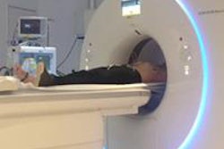
 A 28-year-old woman who was referred for CCTA because of paroxysmal arrhythmia. The examination was
 performed during free breathing with a tube voltage of 80 kVp at a mean heart rate of 51 bpm, resulting in a dose length product of only 24 mGycm. All images courtesy of Dr. Bernhard Bischoff.
CCTA is usually performed during breath-holding to reduce motion artifacts caused by respiration, but some patients are not able to follow the breathing commands adequately due to deafness, hearing impairment, agitation, or pulmonary diseases.
"In these patients, applying standard CCTA scan protocols results in major stair-step artifacts, which often hamper a sufficient evaluation of the coronary arteries. Therefore, it would be desirable to have a scan mode enabling CCTA during free breathing," noted Bischoff and colleagues.
So far, the possibility of performing CCTA during free breathing has only been shown for 320-slice CT units, which allow for acquiring the entire heart without movement of the CT table due to a large z-axis-coverage of 16 cm, they wrote. In most single-source CT systems, the maximum pitch value allowing for gapless image acquisition is approximately 1.5, but in second generation dual-source CT (DSCT) units, the second x-ray tube and detector allow for pitch values of more than 3.0 with a table speed of up to 458 mm/s. This high-pitch scan mode allows for accurate evaluation of the aorta, the pulmonary arteries, and the lung during free breathing in adults and children.
The Munich team evaluated the potential of high-pitch CCTA in free-breathing patients when compared with breath-holding patients in 40 patients (20 breath-holding and 20 free-breathing patients) with a heart rate of 60 bpm or below. Mean heart rate during image acquisition was 52 ±5 bpm in both groups. The mean age of the breath-holding group was 57 ± 18, and 13 (65%) were men, while the mean age of the free breath patients was 55 ± 18, and 11 (55%) were men.
 Dr. Bernhard Bischoff, from the Institute for Clinical Radiology at Ludwig-Maximilians-University Hospital Munich.
Patients with a heart rate above 60 bpm during free breathing were administered up to 20 mg of metoprololtartrate intravenously. Patients with a systolic blood pressure of at least 100 mmHg were administered nitrates sublingually. All examinations were performed on a second generation DSCT system (Somatom Definition Flash, Siemens Healthcare). After acquiring a scout image, CT angiography was planned to cover the entire heart, and initiation of CCTA was timed using bolus tracking.
There was no significant difference in mean image quality, slightly favoring image acquisition during breath-holding. The mean image quality score 3.76 ± 0.32 in breath-holding patients compared with 3.61 ± 0.45 in free-breathing patients (p = 0.411). Due to a smaller amount of injected contrast medium, there was a trend for signal intensity to be slightly lower in free-breathing patients, but this was not statistically significant (435 ± 123 HU vs. 473 ± 117 HU; p = 0.648), the authors explained.
When applying the high-pitch scan mode in DSCT, scan initiation is planned to commence at a certain time point of the RR-interval using prospective ECG-triggering to allow for an image acquisition during diastole with low cardiac motion. Due to low heart rate variability, the intended time point of scan initiation was achieved in a majority of patients without a significant difference between both study groups, they added.
Aside from potential reduction of administered contrast medium, free breathing CCTA might have another benefit. Often a reduced opacification of evaluated vessels is observed, especially after deep inspiration, and this might be explained by a reduced intrathoracic pressure (Valsalva effect), resulting in an increased systemic venous backflow especially in the inferior vena cava with stronger dilution of contrast enhanced blood from the superior vena cava and therefore lower contrast opacification.
"Performing CCTA in free breathing may be a strategy to avoid the Valsalva effect. Since we administered a different amount of contrast medium in both study groups, this hypothesis could not be tested in our study," wrote Bischoff et al.
Also, this was a feasibility study using a relatively small number of patients, and no significant difference in image quality was found between the two groups, they cautioned. Another limitation of this study was the restriction to patients with low heart rates. The applied high pitch scan mode acquires image data of the entire heart during a single diastolic phase of the cardiac cycle.
In patients with low heart rates, the diastolic phase is long enough to allow for image acquisition during low cardiac motion. In patients with higher heart rates, (e.g., more than 60 to 65 bpm), the diastolic phase with low cardiac motion gets shorter, which may result in major motion artifacts when using a high pitch scan mode.
Therefore, the high pitch scan mode in coronary CT angiography should only be used in patients with a low and stable heart rate, although many patients who are not able to hold their breath adequately present with higher heart rates and therefore might not be examined using a high pitch scan mode, they concluded.