Kilovoltage (kV) adaptation can improve CT image quality, cut radiation dose, and lower the required contrast volume, but not all at once, plus it's vital to make a choice, according to Dutch researchers from a top European center of excellence.
KV is not a fixed parameter, kV change requires milliampere-second (mAs) and window adaptation, and it's crucial to keep artifacts in mind, noted Marcel Dijkshoorn, research CT technologist, and colleagues from the department of radiology, Erasmus MC, University of Rotterdam in the Netherlands.
"Kilovoltage is known to be very important in managing radiation dose and contrast in CT. In clinical practice, kilovoltage adaptation is complicated and often not applied within a specific protocol," the authors stated in an e-poster presentation at RSNA 2012 in Chicago, adding that it's necessary to understand kV in standard scan protocols and adapt kV in nonstandard individual patients and cases.
The x-ray beam used for CT scanning has two components. Firstly, millamperes controls the amount of photons, and variation will mainly affect image noise. Secondly, kV controls the energy of the x-ray photons, and variation will affect contrast and noise, they explained.
A decrease of kV settings at equal mAs settings will reduce dose, but dose reduction is not linear compared with mAs change. Also, the noise increase will depend on object size due to the reduced penetration strength of x-rays. The increase in contrast is limited when the Compton effect is dominant (e.g., in the case of a nonenhanced soft-tissue scan), while the increase in contrast is strong when the photoelectric effect is dominant (e.g., in the case of a CT angiography scan), the researchers wrote.
In daily practice, lowering kV can improve image quality by gaining a better contrast-to-noise ratio (CNR) in bone and iodine contrast studies. Also, it can reduce radiation does by gaining an equal CNR in bone and iodine contrast studies, although a small increase in mAs can be achieved with a factor depending on patient size and tissue of interest.
"Lowering kV in noncontrast parenchyma studies will have little benefits," the authors stated. "Lowering kV in large patients can be counterproductive due to strong noise increase or maximum mAs limitations of the system."
In pediatric contrast-enhanced CT, low kV can help to minimize dose by an extra strong contrast increase due to the patients' smaller body size. However, beam hardening is also increased at extra low kV, and to reduce artifacts, it's essential to keep the cabling away from the body part of interest, they continued. Beam hardening artifacts can also arise from undiluted contrast in the subclavian vein, vena cava superior, and right atrium/ventricle. In children, beam hardening artifacts are stronger at low kV, and more of the anatomy needs covering than in adults, while extra care must be taken to avoid these artifacts by correct saline or mixed bolus chasers.
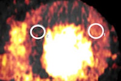
Editor's note: The CT image used to illustrate this article on our home page was provided by Philips Healthcare.