Whole-body CT might be a poor choice for screening asymptomatic individuals, but it has proved its worth in emergency departments, detecting significant and sometimes unexpected injuries in seriously injured patients.
Delayed scans can also be helpful in the emergency setting. The question is: Would the routine acquisition of a delayed scan five minutes after contrast lead to better diagnoses of trauma patients? Delayed CT is known to improve visualization and diagnostic confidence in some cases, but is it worth the additional time and radiation dose? If so, which trauma patients are the best candidates?
It pays sometimes but not always to acquire the extra scan, say researchers from Italy, who reviewed images from hundreds of cases of organ injuries in which patients received delayed scans in addition to arterial and venous-phase imaging.
"Whole-body MDCT acquisition protocols allow simultaneous evaluation of vascular and solid-organ injuries, but [CT] doesn't opacify renal collecting systems: ureters and bladders," said Dr. Alessandro Lemos from Foundation IRCCS Policlinico and the University of Milan in Italy. Delayed CT of the abdomen not only allows time for adequate opacification of the urinary collecting system, "it allows detection of active extravasation and characterization of splenic injuries, especially pseudoaneurysm versus active bleeding," Lemos said in a presentation at the 2008 RSNA meeting in Chicago.
Lemos and his colleague Dr. Pietro Biondetti retrospectively evaluated the utility of delayed CT of the abdomen and pelvis and its potential role as selective acquisition integrated into whole-body CT. The inclusion criteria included patients with solid organ injury, bowel or mesenteric injury, free fluid, active contrast extravasation, or pelvic fracture. Patients were excluded if no delayed-phase imaging was performed, or if there was no automatic modulation of the radiation dose, for example in obese patients.
"Delayed scans were considered useful when they aided in characterizing initial CT findings, identifying findings not present at initial CT, or increasing reader confidence with regard to initial CT findings," Lemos said. A normal approximation of the binomial distribution was used to obtain confidence intervals.
The researchers examined a total of 562 patients (397 men, 165 women; mean age, 40.2 years; range, 18-91) who were given whole-body CT with five-minute delayed-phase images over a period of two years, identifying injuries in 96 patients.
"We acquired a complete arterial phase, and after that we acquired a venous phase, and then, depending on the scenario, a delayed phase," Lemos said. "Two radiologists blinded to the initial CT scan interpretation reviewed these cases to determine the utility of delayed scans. The reviewers were also asked to record the quality of the delayed scans as being either diagnostic or nondiagnostic on the basis of the organ or area of interest."
 |
| A 45-year-old man after a car collision (left). Initial axial CT performed after splenic artery embolization (white arrow), shows a round area of increased attenuation in the spleen (black arrow).This finding was believed to represent active bleeding, but organ injury was not visible. Five-minute delayed scan (right) demonstrated intraperitoneal fluid, hematoma, and contrast material extravasation, confirming that the finding was secondary to delayed splenic rupture. The patient subsequently underwent splenectomy. All images and data courtesy of Dr. Alessandro Lemos. |
The researchers found the delayed scans to be helpful in 20% (9 of 45) of patients with solid organ injuries, 16% (2 of 12) of patients with bowel or mesenteric injuries, 12% (3 of 25) of patients with pelvic fractures, and 14% (2 of 14) of patients with free fluid only.
 |
| Although delayed CT scanning was helpful in only 2.8% of patients overall, it was helpful in 20% of solid organ injuries and in 16% of bowel or mesenteric injuries. In patients with an initial CT scan suggesting injury, delayed CT was helpful in 16.6% of cases. |
Overall, delayed CT was useful in just 2.8% (16 of 562) of all patients (95% confidence interval, 1.2-3.5) referred for evaluation following trauma as part of a whole-body CT protocol, Lemos said. But its utility rose to 16.6% (16 of 96; 95% confidence interval, 7.5-19) in patients with injuries detected or those suspected of having injuries after initial CT.
 |
| A 35-year-old woman after a motorbike collision with pelvic impact. Initial axial CT scan (left) shows high-attenuation perihepatic fluid (arrow). Five-minute delayed scan (right) demonstrates bladder dome rupture with clot covering (arrow) and contrast material extravasation in the intraperitoneal space (arrowheads). The patient subsequently underwent bladder repair. |
"Delayed phase was most useful in solid organ injuries -- in 20% -- whereas the utility in patients with suspected injury after CT was 16.6%, and there was an increase in confidence intervals," he said.
 |
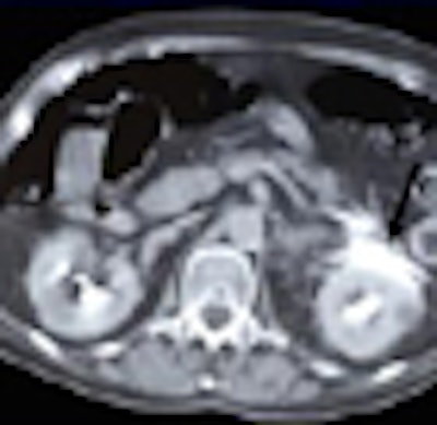
| A 52-year-old man after a motor vehicle collision. Initial axial CT scan (left) shows free fluid (arrow). This finding was suspicious for pancreatic injury. Five-minute delayed scan (right) demonstrates rupture of the upper pole of the left kidney associated with contrast material extravasation (arrow). The patient was treated conservatively. |
"Although the integration of delayed phase into whole-body CT trauma imaging allows additional investigations, selective rather than routine acquisition is recommended to avoid overexposure and data explosion," Lemos said. "Delayed CT scan should be acquired in patients with injury or suspected injury after initial CT, particularly when free abdominal fluid is detected."
By Eric Barnes
AuntMinnie.com staff writer
January 21, 2009
Related Reading
Studies address need to reduce CT dose in emergency settings, October 17, 2008
When zoos refuse: Obese patients face shortage of large-capacity scanners, September 24, 2008
CTA assesses traumatic neurovascular injury, October 16, 2006
3D penetrates trauma imaging niche, August 4, 2006
Copyright © 2009 AuntMinnie.com