The radiation dose of pediatric MDCT exams can be reduced by as much as 75% without compromising the ability to detect small lung nodules in young children, according to a study published in the July issue of Academic Radiology.
Researchers from Duke University Medical Center in Durham, NC, found that a panel of three radiologists reported only minor differences in diagnostic accuracy when reviewing pediatric CT chest images performed at three levels of lower tube current (and, thus, lower radiation dose) (Academic Radiology, July 2009, Vol. 16:7, pp. 872-880).
What's more, the Duke researchers derived the findings using a new noise simulation tool that may enable pediatric radiologists to examine the impact of various CT protocols on image quality, without subjecting children to additional CT studies. In a related study, Swedish researchers used a similar simulation tool to find out that pediatric dose reduction needs to be selectively applied, or else diagnostic accuracy may suffer.
Noise simulation
Assessing the effects of different CT scanning protocols on image quality can be a tricky business in children, because it's not ethical to perform repeat scans on the same patients at different tube currents. In addition, the low occurrence of isolated small lung nodules in children would make it difficult to enroll enough patients to conduct a clinical study.
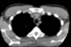
To address this issue, principal investigator Xiang Li, a doctoral student at Duke Advanced Imaging Laboratories, led a research team that used noise simulation software (Noise Addition Tool, GE Healthcare, Chalfont St. Giles, U.K.) and a nodule simulation technique developed at Duke. The researchers applied the techniques to normal chest CT exams of 13 patients ranging in age from 1 to 7 years (median age, 3 years) and in weight from 24.2 to 50.6 lb (11-23 kg), with a median weight of 30.8 pounds (14 kg).
The original normal images were acquired with a 64-slice MDCT scanner (LightSpeed VCT, GE Healthcare) using Duke's size-based pediatric clinical protocols: tube current of 70 to 180 mA, tube potential of 100 or 120 kVp, beam collimation of 20 or 40 mm, a pitch of 1.375, and a slice thickness of 3.75 mm at a 3.75-mm slice interval, or 5-mm thickness at a 5-mm interval.
The noise simulation tool was used to create three sets of the 13 exams at different tube currents: 70 mA, 35 mA, and 17.5 mA. Three copies of each simulated dose reduction were made to enlarge the scale of the study to a total of 117 CT exams.
Researchers used Duke's nodule simulation technique to create small lung nodules 3-5 mm, with peak contrasts of 250 to 550 HU, and added them to the 117 series of images. After dividing the lung volumes in each series into upper, middle, and lower anatomic zones, the researchers added simulated nodules so that each of the three lung zones contained either none or one lung nodule. A total of five nodules were inserted in the nine lung zones of the three copies of each case.
Three pediatric radiologists with different levels of experience independently read all 117 series. These were displayed in random order on a clinical workstation (Advantage Workstation, GE Healthcare) in a controlled viewing environment. Each radiologist rated the presence of a nodule in each lung zone on a 0 to 100 scale, and identified the location of a possible nodule. The results are indicated below, as measured by the area under the receiver operator characteristics (ROC) curve:
Radiologist accuracy at various simulated tube currents
|
||||||||||||||||||||||||
The authors acknowledged that while the average decrease in accuracy in detecting lung nodules was only 3% and interobserver variability was only 2%, whether this would be clinically significant was beyond the scope of their study and could only be validated by a large-scale, long-term, and probably impractical clinical study. Li and colleagues also acknowledge that because the radiologists knew they were only looking for lung nodules, their performance may have been positively impacted because they did not have to look for any other disease.
However, in spite of the acknowledged limitations of the study, the authors suggested that radiation dose for pediatric CT exams can be lowered without comprising the ability to identify lung nodules in young children.
Swedish study
In a related study, researchers from Sahlgrenska University Hospital and radiologists from the Queen Silvia Children's Hospital, both located in Göteborg, also used noise simulation to investigate the effects of tube current on pediatric CT studies, this time brain exams. They described their methodology in the April issue of the British Journal of Radiology (2009, Vol. 82, pp. 313-320).
A research team led by Dr. Kerstin Ledenius selected brain CT exams that had been performed on patients ranging in age from newborn to 15 years over a four-month period of time. These used the institution's standard head protocols, which have already been optimized for the age and body size of patients to keep dose low.
The researchers then used GE noise simulation software to simulate exams performed at 50/70 mAs for patients 11 to 14 years of age; 40/60 mAs for patients older than 14 and ages 1 to 10; and 30/50 mAs for patients younger than 1 year. Three readers were asked to review the images to evaluate image quality.
Ledenius and colleagues found that the impact of dose reduction varied based on patient age. For patients between the ages of 1 and 10, the institution could reduce dose by 20% while preserving image quality. For patients older than 10, reducing mAs resulted in unacceptable image quality, and the institution's standard scanning protocols were maintained.
For patients younger than 6 months, the group found that the hospital's existing protocols were producing unacceptable images, and that dose should be increased slightly. A similar finding was reached for patients between the ages of 6 months and 1 year.
By Cynthia E. Keen
AuntMinnie.com staff writer
June 18, 2009
Related Reading
Color coded CT protocols help reduce pediatric radiation dose, June 4, 2009
SPR news: Rads must take lead in reducing pediatric CT dose, April 23, 2009
ARRS study: Child's body shape can reduce CT dose, April 23, 2009
Pediatric CT dose drops with breast shields plus tube modulation, February 26, 2008
Copyright © 2009 AuntMinnie.com