For detecting aortic emergencies, CT angiography (CTA) is the modality of choice and enables clinical teams to decide whether elective, urgent, or emergent treatment is necessary, while endovascular aortic repair (EVAR) and surgical repair are the recommended therapies, according to Austrian researchers.
Aortic emergencies are a challenge for physicians, because they are different clinical entities with only one thing in common: If not diagnosed and managed in due time, they are life-threatening, noted Dr. Esther Voitle from the Institute for Diagnostic and Interventional Radiology, Academic Teaching Hospital in Feldkirch, Austria, and colleagues.
"Compared to myocardial infarction and stroke, aortic pathologies are relatively rare," they wrote. "A rapid diagnosis and treatment decision is therefore necessary to decrease mortality. CTA has become the best modality for the diagnosis and treatment planning of aortic pathologies. Treatment is performed by either an open surgical approach or endovascular stent graft implantation (endovascular aortic repair, EVAR)."
The researchers sought to demonstrate the various presentations of acute aortic pathology and to present diagnostic and therapeutic approaches (Insights into Imaging, 1 February 2015).
"CT is unsurpassed in its ability to rapidly detect aortic pathology, sometimes already in noncontrasted scans (e.g., aneurysm rupture or intramural hematoma)," they wrote. "However, noncontrast scans are not always necessary for diagnosis; multiphasic scans multiply the total radiation burden."
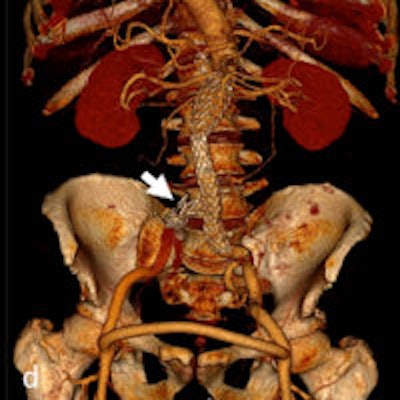
 Ruptured abdominal aortic aneurysm with stent graft treatment. The 72-year-old man presented with left-sided abdominal pain and hypotension. CTA demonstrated a ruptured infrarenal aortic aneurysm on coronal and axial sections (a-b). The patient was treated with aorto-uni-iliac stent graft implantation (d) to the left, a crossover bypass, and placement of an occluder (arrow) in the right common iliac artery. Follow-up CTA (c) demonstrated complete exclusion of the large aneurysm. All images courtesy of Dr. Esther Voitle, from Insights into Imaging.
CTA is usually reserved for true arterial phase CT. However, all aortic pathologies or associated pathologies are easily depicted in both the arterial and delayed phases. Delayed phases are often included in an imaging protocol -- in addition to CTA -- to clearly depict organ malperfusion (in dissections) or identify additional venous or organ bleeding.
"Fast imaging, quick image reconstruction, and image transfer to the workstation and fast (3D) image processing are the main advantages of modern CT systems in emergency radiology," they added.
Also, 3D vascular reconstruction with vascular segmentation and centerline reconstructions are often helpful in aortic measurements for stent graft planning, which is easier to perform in the arterial phase than in the venous phase.
"The drawbacks of CT are the use of contrast media and ionizing radiation," they wrote. "Contrast media may pose a threat of worsening of renal function in patients with chronic renal insufficiency, although CTA imaging of the whole aorta is possible with as little as 40-50 mL of contrast with 64-slice systems."
 Ruptured type B dissection with stent graft implantation. An 83-year-old patient with ruptured type B dissection in parasagittal reformations demonstrating intimal tear (a) and hematothorax (a, b). Stent graft implantation (c) (partially covering the left subclavian artery) and exclusion of the dissection after expansion of the stent graft (d).
Voitle and colleagues outline several different aortic pathologies in their article, including the following:
Traumatic aortic injury: A life-threatening consequence of major blunt thoracic trauma, with high early mortality if relevant trauma is untreated. It has to be excluded after a relevant trauma, especially when associated with hypotension, fractures -- including those of the spine, scapula long bone, or pelvis -- pulmonary contusion, and hematothorax.
Acute aortic syndrome: Comprises aortic dissection (AD), intramural hematoma (IMH), and penetrating atherosclerotic ulcer (PAU).
Aortic dissection: An entrance tear from the lumen to the media that allows blood flow into the vascular wall and therefore creates a false lumen, usually with additional communication tears between the old "true" and the new "false" lumen.
Ruptured aortic aneurysm: The diagnosis of which depends on measurement of the vascular diameter.
Mycotic or infected aneurysms: These rare but life-threatening aneurysms are usually detected when they become symptomatic. They have an atypical morphological presentation, as saccular aneurysms, eccentric aneurysms, or pseudoaneurysms, but they may also present as grotesque fusiform aneurysms.
Aortoenteric or aortoduodenal fistulae: Rare but often fatal with late complications of open repair (after graft implantation) or aneurysm infection. The clinical presentation is often with massive gastrointestinal bleeding, starting with herald bleeds and leading to severe bleeding episodes with exsanguination.
Iatrogenic aortic injury: May be caused by direct external injury from surgical procedures or an endovascular approach.
The researchers found CTA is reliable in diagnosing and grading aortic trauma, measuring aortic diameter in aortic aneurysms, and detecting vascular wall pathology in acute aortic syndrome and aortic inflammation. CTA also allows for planning the optimal therapeutic approach. In addition, stent graft implantation and/or an open surgical approach can address vascular wall pathology and exclude aortic aneurysms, they added.