Scientists in France have developed a new type of ultrasound that can produce 3D videos at thousands of frames per second. The 3D ultrafast ultrasound builds on the established technique of 2D ultrafast ultrasound, and has already enabled its creators to witness blood flowing through the chambers of a human heart in real-time.
Two-dimensional ultrafast ultrasound imaging was invented in 1999 by Mathias Fink (ESPCI professor and director of the Institut Langevin of ESPCI ParisTech) and Mickael Tanter (director of research at INSERM [DR1] and a medical physicist at Université Paris VII) through developments in the software and hardware used to process ultrasound images.
It allowed the scientists to observe the mechanical shear waves that propagate rapidly through the body at several meters per second -- and thereby opened up a kind of human-body "seismology" that could map the elastic properties of soft tissue. At least one commercial ultrasound scanner now delivers ultrafast 2D images, and many new applications have come along: mapping the flow of blood at high sensitivity, for example, or quantitatively characterizing tumors.
','dvPres', 'clsBtn', 'true' );" >
For some applications, however, 2D is not enough. Shear waves propagating through the body have components in 3D, and so assessing them in just 2D relies on certain assumptions that can be inaccurate. This is an important consideration if, for example, a doctor is attempting to determine a precise location in an irregularly beating heart to site a pacemaker.
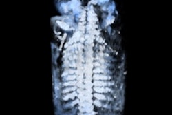
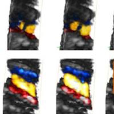
Now, Tanter and colleagues at ESPCI Paris Tech (a chemistry and physics engineering college run by the city of Paris)and other French institutions have extended the technique of 2D ultrafast ultrasound imaging to 3D. The researchers used a probe with a matrix of 1,024 elements to emit plane or diverging ultrasound waves, and processed the backscattered signals on a computer with a special graphics processing unit. Unlike currently available 3D ultrasound, which generates just 10 to 20 volumes per second, their 3D ultrasound could generate thousands of volumes per second (Physics in Medicine and Biology, 10 September 2014, Vol. 59:19, L1-L13).
The researchers tested their ultrafast ultrasound by imaging shear waves in the human body in 3D in real-time, which in future could allow for better characterization of lesions in the breast, liver, or prostate. They also imaged cardiac blood flow in 3D, potentially enabling better diagnosis of cardiovascular problems and mapping the vasculature of infant organs such as the brain, the kidneys, and the liver.
"To our knowledge, it is the first time that cardiac blood flows were imaged in 3D in real-time," said co-author Mathieu Pernot, PhD.
Pernot, who is INSERM research associate at Institut Langevin in Paris and a former research scientist at Supersonic Imagine, Aix-en-Provence, France, believes that there is still some way to go before 3D ultrafast ultrasound can join its 2D counterpart in hospitals.
"This imaging platform is a research prototype," he said. "A lot of work must be performed before it can be used in clinical practice -- for example, its size and cost need to be reduced. Technology is progressing very fast, however, and we are confident that 3D ultrafast imaging will become widely available in the near future."
© IOP Publishing Limited. Republished with permission from medicalphysicsweb, a community website covering fundamental research and emerging technologies in medical imaging and radiation therapy.