Aligning the lower limbs for orthopedic surgery is a tough job now made easier with the use of an augmented realty C-Arm technique, said researchers from Munich, Germany, in the November edition of International Journal of Computer Assisted Radiology and Surgery.
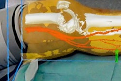
The team used a mobile C-arm system augmented with a vide camera to create a panoramic image consisting of hip, knee, and ankle x-rays in a bid to replace the dose-inefficient and time-consuming fluoroscopy examination used currently. The results based on 25 cadaver legs showed a strong positive correlation between the C-arm and ground-truth CT data acquired to test the process.
"We showed that only a few x-ray images are sufficient to measure a clinically relevant mechanical axis deviation," wrote lead author Pascal Fallavollita, PhD, former chair for computer-aided medical procedures and augmented reality at the Technical University of Munich and Munich University Clinic, in an email to AuntMinnieEurope.com. He recently moved to the University of Ottawa in Canada and is now an assistant professor.
Easier lower-limb alignment
The C-arm-based augmented reality technology system offers accurate mechanical axis deviation measurements for lower-limb alignment, Fallavollita and colleagues reported.
 Pascal Fallavollita, PhD.
The C-arm system combines an x-ray and video overlay in real-time, and by visualizing a marker pattern, generates an x-ray panorama, they explained (IJCARS, November 2016, Vol. 11:11, pp. 2111-2117).
The surgical procedure known as high tibial osteotomy was developed in 1961, and it became popular for correcting deformities and reducing pain in the treatment of osteoarthritic knee, they wrote.
The idea is to redistribute the weight-bearing load from the arthritic portion of the noninvolved articular cartilage portion of the knee.
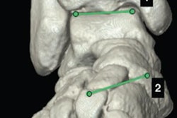
But it's not an easy job. Evaluation of the alignment before surgery requires assessment of the frontal plane mechanical axis of the entire limb and not just the bones. To do this, the mechanical axis deviation (MAD) measures the distance from the knee joint center to the line connecting the joint centers of the hip and ankle, the study team explained.
In normal individuals, the mechanical axis of the lower extremity lies from 1 mm to 15 mm medial (average 8 mm ± 7 mm) to the knee joint center, the group wrote.
However, determining the exact MAD intraoperatively is a difficult clinical problem such that no good way has been found to do it so far. Clinicians have used techniques like the electrocautery cord, alignment rods, or axis boards to solve the problem, and studies have shown no significant difference in their measurements.
But this type of assessment is only momentary, and all of them are prone to technical mistakes, resulting in large numbers of x-ray acquisitions until the alignment can be closely estimated. The x-ray images must show the cord, board, or rod centered on the hip, knee, and ankles in order to perform the MAD measurement subsequently.
3 images, lower dose
In this study, the authors used C-arm technology to create a panorama image consisting of hip, knee, and ankle x-rays together.
"The purpose of this paper was to evaluate whether an augmented reality technology can provide accurate, efficient, and reliable intraoperative alignment control of the MAD," Fallavollita and colleagues wrote.
The solution offers the advantage of needing just three x-ray images, and the anatomy need not be centered within the x-rays. The researchers assessed this technique in a preclinical study of 25 cadaver legs with five clinicians of various experience levels.
Using the C-arm, the acquired x-ray image is registered together with the video image without any further calibration or registration needed intraoperatively, according to the authors.
Due to the mirror construction and a one-time calibration of the device, the acquired x-ray image is registered with the video image without any further calibration or registration. Users can acquire real-time pose information by placing a planar marker underneath the limb while it is detected and tracked with the video camera.
"In total, a couple of minutes are required to acquire the x-ray images and calculate the intraoperative mechanical axis deviation measurement," Fallavollita wrote in his email.
Multiple benefits of this approach have been shown in applications such as the six-degree-of-freedom (DOF) kinematic modeling of a C-arm, video-guided calibration of a C-arm, parallax-free panorama generation for total knee arthroplasty surgery, and multimodal perceptual visualization, they noted.
This study used 25 human cadaver legs for validation, all with random varus or valgus deformations. Five clinicians with different surgical experience levels performed experiments aimed at achieving acceptable mechanical axis deviation. Each observer manually determined the anatomical landmarks defining the mechanical axis and knee center using the ground-truth CT images and x-ray images.
To do so they assessed the applicability of C-arm technology to ground-truth CT. A t test, Pearson's correlation, and analysis of variance were used to determine statistical significance.
Close correlation with CT
The results showed a strong positive correlation between the C-arm with CT results, with the t test revealing 13.71 mm ± 11.57 mm for the CT data, compared with 13.19 mm ± 10.55 mm when using the C-arm data, with a Pearson's correlation coefficient R value of 0.979.
An analysis of variance yielded a p-value of 0.011, showing that differences in the expertise of the five operators were not significant with regard to the system used to assess mechanical access deviation.
"Our investigation demonstrated that the augmented reality technology provides accurate mechanical axis deviation measurements for lower limb alignment," the authors concluded.
The C-arm system combines an x-ray and video overlay in real-time, generating an x-ray panorama by visualizing a marker pattern the study team wrote. MAD errors are well within patient tolerances.
Moreover, the clinician has the option of selecting more or less than three images to create the panorama depending on the procedure and the targeted anatomy -- in this experiment consisting of the hip, knee, and ankle -- with the key advantage that the anatomy need not be centered within the images, they wrote.
Going forward, the user interface needs improvement to make it more intuitive for the clinicians when reading the measurements for the mechanical axis deviation, Fallavollita noted. Before the system is adopted for daily use, a new study will integrate all of the elements into real-time clinical workflow.
"Our next focus should be on a final preclinical study that investigates all aspects of the surgical workflow and how our novel augmented reality technology affects the surgical team during intervention," he stated. "This means measuring total radiation exposure and procedure time compared to traditional imaging solutions such as mobile C-arms."