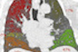
A 3D algorithm can utilize PET/CT images to map the movement of oxygen and CO2 in the lungs, potentially facilitating better treatment for patients with serious lung diseases, according to a recent article published online in Respiratory Physiology & Neurobiology (10 November 2015).
Detailed knowledge about the movement of oxygen and CO2 in the lungs is important for patients with serious lung disease. The new technique maps that movement in detail by essentially creating a 3D map of how and where the CO2 and oxygen transfers take place, according to lead study author Dr. Troels Johansen, a doctoral student from Aarhus University in Denmark.
As part of this doctoral project, Johansen collaborated with Harvard Medical School to develop a mathematical model that provides the basis for the 3D images, which are developed from PET/CT scans.
In lung cancer patients, for example, the model can help predict the consequences of removing part of the lung by surgery. It will also help doctors predict which chronic obstructive pulmonary disease (COPD) patients will benefit from surgery and which will not.
In addition to enabling doctors to foresee the consequences of high-risk lung operations, the model will also provide important new basic knowledge about oxygen and CO2 transfer in healthy individuals and those with serious lung disease, the authors said.