While breast MRI is highly sensitive for breast cancer, its specificity can be a little lackluster. Fusing FDG-PET and breast MRI can help, however, Brazilian researchers reported in an article published online on 27 May in the European Journal of Radiology.
In a study of 76 lesions, researchers from Hospital A.C. Camargo in São Paulo found that PET/MRI fusion yielded sensitivity of nearly 90% and specificity of more than 75% for all lesions. For mass lesions larger than 10 mm, sensitivity climbed to approximately 96% and specificity topped 83%.
"We believe that this new methodology has the potential to help more effectively in the management of breast lesions in the near future, with the incorporation of dedicated PET/MRI devices, improvement of the spatial resolution of PET scanners, introduction of new radiotracers and correlation with functional MRI data," wrote a team led by Dr. Almir Bitencourt.
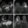
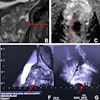
To see if the functional information provided via FDG-PET could improve the specificity of MRI, the research team selected 60 patients who had suspicious breast lesions on MRI. These patients received a PET/CT scan in prone position for evaluation of the breasts.
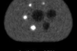
After areas with increased FDG concentration relative to normal parenchyma had been observed, a radiologist experienced with breast MRI performed fusion of PET and MRI using version 4 of the Aquarius advanced visualization software (TeraRecon). The PET/MRI study was considered to be positive if there was topographic correspondence between the area of increased concentration of FDG on PET/CT and the suspicious lesion identified on MRI, according to the researchers.
The 60 patients had 76 lesions included 64 mass lesions and 12 nonmass lesions with a mean diameter on MRI of 29.6 ± 19.2 mm and a range of 6-94 mm. PET/CT revealed increased metabolic activity on 57 (75%) lesions with a mean maximum standard uptake value (SUV) of 5.7 ± 5.0 and a range of 0.8 to 23.1. PET/MRI fusion findings were compared with histopathological results.
| PET/MRI fusion sensitivity | |||
| Sensitivity | Specificity | Accuracy | |
| All lesions | 89.8% | 76.5% | 86.8% |
| Lesions larger than 10 mm | 95.8% | 83.3% | 93.3% |
The results showed that PET can improve the specificity of MRI, particularly for masses ≥ 10 mm, according to the researchers.
"When we evaluated only the BIRADS 4 lesions on MRI, which is the group with most diagnostic divergence, we observed that increased [FDG] uptake on PET was statistically significantly associated with malignancy," they noted. "However, the number of false-negative results does not yet allow us to waive the histological evaluation on suspicious breast lesions on MRI without abnormal glycolytic metabolism on [FDG] PET."
Adding FDG-PET to the evaluation of suspicious breast lesions on MRI allows "for the identification of most aggressive tumors at histological analysis," they concluded.