VIENNA - Breast MRI computer-aided detection (CAD) technology can effectively distinguish between benign and malignant lesions, according to a presentation at the 2010 European Congress of Radiology (ECR).
A German research team found that an automatic breast MRI CAD system produced sensitivity of 96.8% and specificity of 78.9% in a study of 101 contrast-enhancing breast lesions.
"The analyzed novel CAD system may be a promising tool as a second reader for the diagnosis of MR mammography in clinical routine," said presenter Dr. Diane Renz of Charité Medical University in Berlin.
Although breast MRI CAD systems have been available for several years, use of the technology as a second reader remains controversial. To see if a CAD system that automatically considers both dynamic and morphological information could demonstrate value, the Charité researchers enrolled 80 patients (mean age, 51.4) with 105 histologically-proven mass-like lesions (including 63 cancers) in their study.
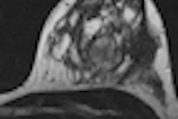
MRI scans were performed on a 1.5-tesla scanner with T1-weighted dynamic sequences and 0.2 mmol/kg body weight of gadolinium-diethylenetriaminepentaacetic acid (Gd-DTPA) contrast. The study team then applied the Breast MRI Carebox software (Bracco Imaging, Milan, Italy), which offers automated analysis without any action by the user.
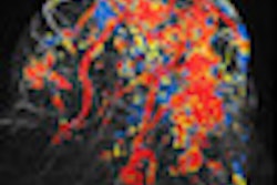
The system automatically performs motion correction (using a 3D elastic registration algorithm), 4D threshold-based image segmentation with detection of lesions, calculation of several dynamic and morphological parameters, and lesion classification. Via color-coded parametric maps, the software summarizes the morphological and dynamic features for characterizing the lesion, Renz said. Five colors are used, ranging from green (benign) to red (malignant).
The CAD system concludes its analysis by estimating the probability of malignancy via a morphodynamic index (MDI), which ranges from 0 to 100%, she said.
The CAD system was able to detect 101 of 105 enhancing masses. Four enhancing masses were not found by the system, including two fibroadenomas (12 mm and 5 mm), one atypical ductal hyperplasia, and one malignant focus.
In the 101 masses found by the system, CAD provided a mean MDI score of 88.6% for malignant lesions and 39.1% for benign lesions. The difference was statistically significant (p < 0.001).
Using a cutoff MDI score of 50%, CAD turned in sensitivity of 96.8% and specificity of 78.9%, Renz said.
"Correlation analysis with histologic findings reveals that a fully-automatic CAD system seems to have the ability to distinguish between benign and malignant enhancing breast masses," she said.
Renz acknowledged limitations of the system, which is not capable of considering T2-weighted images. This potentially may miss useful additional morphology information available in those sequences, she said. Also, only mass-like lesions were included in the study.
Nonetheless, Renz said, the system shows promise as a second reader.
"Going further, it would be interesting to evaluate the benefit of this method in a clinical setting, [such as] analyzing the diagnostic accuracy of a radiologist without CAD and afterward with the assistance of the automatic method," she said.
By Erik L. Ridley
AuntMinnie.com staff writer
March 8, 2010
Related Reading
CAD enhances breast MRI specificity, September 17, 2009
Breast MRI CAD algorithm predicts cancer metastasis, February 6, 2009
Reimbursement crunch throttles breast MRI CAD users, April 8, 2008
Breast MRI CAD doesn't improve accuracy due to poor DCIS detection, February 18, 2008
Copyright © 2010 AuntMinnie.com