Dual-energy cardiac CT can readily distinguish cardiac sarcoidosis from other kinds of cardiac hypertrophy, eliminating the need for cardiac MRI, according to researchers from France.
Researchers from Hopitaux Universitaires Henri-Mondor in Créteil, Paris, used dual-energy CT to examine patients with confirmed sarcoidosis, and compared the results with patients with other types of cardiac hypertrophy and a control group. The dual-energy CT protocol was able to distinguish the cardiac sarcoidosis cases with high per-patient accuracy.
"This is the first study to look at iodine concentration using dual-energy CT in patients with cardiac amyloidosis and distinguish them from other cases of hypertrophy with 99% accuracy," said Dr. Virgile Chevance in a presentation at the RSNA 2016 meeting.
Replacing heart muscle
Cardiac amyloidosis occurs when amyloid deposits begin to replace normal heart muscle. Cardiac amyloidosis, the most common type of cardiac hypertrophy, can affect the heart's electrical conduction system, potentially leading to abnormal heartbeats and faulty heart signals. The condition is typically diagnosed with cardiac MRI, but the modality cannot be used in most patients with cardiac devices, or other contraindications to MRI.
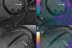
Two recent studies have shown an increase in CT attenuation values after contrast injection, in the setting of rising extracellular volumes in patients with cardiac amyloidosis. CT allows clinicians to assess myocardial iodine concentrations, Chevance said. These results suggest amyloidosis may be readily detected on CT images, obviating the need for MRI and its limitations.
"We propose to compare myocardial iodine attenuation using dual-energy CT in patients with cardiac amyloidosis in comparison to patients with other causes of cardiac hypertrophy and a group of control subjects," he noted.
The prospective study included 22 patients with cardiac amyloidosis, 13 patients with nonamyloid cardiomyopathies other causes of hypertrophy, and 10 control subjects. All patients underwent multienergy cardiac CT with Gemstone spectral technology (Discovery CT750 HD, GE Healthcare).
The investigators acquired a noncontrast phase, an arterial phase, and a delayed-enhancement phase five minutes after injection of contrast medium (iomeprol 1.5 mL/kg.). The investigators used a dedicated workstation (Advantage Workstation 4.6, GE Healthcare) to calculate interventricular septum thickness and myocardial iodine concentration on enhanced images. The latter group underwent imaging for suspected thrombosis in the left atrial appendage, he said.
Clear differences in amyloidosis patients
The researchers discovered that dual-energy CT of iodine concentration revealed a number of differences between the patient groups. For example, interventricular septum thickness was higher on a statistically significant basis in both cardiac amyloidosis and nonamyloid hypertrophic cardiomyopathies (HCM) than in control patients, the authors explained.
Also, cardiac amyloidosis patients exhibited higher iodine concentration within the myocardium than both hypertrophic cardiomyopathy patients and control patients, the authors explained, also results that were statistically significant.
| Dual-energy CT of cardiac amyloidosis | ||||
| Parameter | Cardiac amyloidosis | Nonamyloid hypertrophic cardiomyopathies | Control group | P value |
| Intraventricular septal thickness | 17 ± 4 mm | 15 ± 3 mm | 10 ± 1 mm | p < 0.01 |
| Myocardial iodine concentration | 2.69 ± 0.6 mg.mL-1 | 1.84 ± 0.5 mg.mL-1 (p < 0.01) |
1.93 ± 0.4 mg.mL-1 | p < 0.005 |
The area under curve of mean iodine concentration for diagnosing cardiac amyloidosis patients compared with hypertrophic cardiomyopathy patients was 0.86 (0.73-1.0; p = 0.001), they noted. With a cutoff value of 2.22 mg.mL-1, the sensitivity and specificity of iodine concentration for diagnosis of cardiac amyloidosis were 82% and 77%, respectively.
"The patients with sarcoidosis had elevated enzymes including troponins compared with control patients, and the patients with sarcoidosis had an increase in the intraventricular septum thickness compared to control patients," Chevance said. "Regarding the iodine concentration ratio there was a difference between the groups at the arterial phase, and at the five-minute [delayed] acquisition the patients with amyloidosis had an increase in the iodine concentration ratio compared to other patients."
"There are several advantages of cardiac CT, like exam time, accessibility, and access, and in particular it is feasible in patients with cardiac devices," he continued, adding that on the other hand, triple-phase CT carries a radiation burden. Another limitation of the modality was the lack of correlation with MRI results because patients who were not contraindicated for MRI had already had an exam.
"Most patients had cardiac devices," he said. "We did the study so we could begin to include patients with cardiac devices."
In response to a question from the RSNA audience, Chevance said he believes the mechanism of higher attenuation in cardiac amyloidosis patients relates to the additional myocardial contrast.
"I think it's just an increase of the extracellular volume so that the contrast was pooled inside the myocardium," he said.