Initially a technology used only by early adopters, 3D/4D ultrasound has indeed come of age, buoyed by mounting evidence of its diagnostic utility in a variety of clinical applications. Today, 3D/4D ultrasound is increasingly being used by healthcare institutions around the world.
In its early years, the diagnostic evidence for 3D ultrasound was not strong enough to convert scanning procedures from 2D to 3D. However, the seeds of change were planted. As I discussed in a 2013 article for AuntMinnie.com on the coming of age of 3D/4D ultrasound, a number of key trends have driven the proliferation of the technology.
 Isabelle Wegmann Hachette from ContextVision.
Now in 2016, the advantages of 3D/4D ultrasound have become even clearer, thanks to advances in hardware and software. On the hardware end, the continued miniaturization of equipment is leading to more portable and easier-to-use transducers with better image acquisition, higher image resolution, and more data. This improves diagnostic accuracy.
Thanks to 3D ultrasound's more comprehensive image acquisition techniques, you can also now acquire every structure of the examined volume, making reconstruction possible in a multitude of image planes. When you acquire a 2D structure, you might miss something that needs another look with the ultrasound equipment, adding to the cost and time it takes to arrive at a proper diagnosis.
As we make our way into 2017 and beyond, let's revisit the key trends affecting 3D/4D ultrasound's diagnostic value and explore some of the factors driving the acceleration predicted in 2013. We'll then explore how image processing algorithms can make it easier to interpret these studies.
Diagnostic improvements
With 3D, the whole anatomy can be acquired in a single scan for many clinical applications. Volumetric scanning leaves no information behind, ultimately leading to a faster and more accurate diagnosis. At the 2012 International Society of Ultrasound in Obstetrics and Gynecology (ISUOG) annual meeting, Dr. Beryl Benacerraf of Brigham and Women's Hospital said the "volume contains all of the information available and we can display the image in any plane. Identification, positioning, and volume quantification improve diagnostic quality and measurement accuracy with 3D in new ways that 2D has never been capable of."
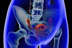
In gynecology, 3D ultrasounds can be helpful in many circumstances, including but not limited to evaluating the relationship of masses in the endometrial cavity, identifying uterine congenital anomalies and a thickened and/or heterogeneous endometrium, and evaluating the location of an intrauterine device and the integrity of the pelvic floor, according to Benacerraf and guidelines found in the 2014 American Institute of Ultrasound in Medicine (AIUM) practice parameter "Ultrasound of the Female Pelvis." Specifically, uterus malformations such as a bicornuate, or heart-shaped, uterus can be easily evaluated with 3D, Benacerraf said at the 2012 ISUOG meeting.
 Uterus shape visualized with 3D thick-slice image (left) and 3D volume (right). All images courtesy of ContextVision.
In reproductive medicine, the ovaries are measured to detect abnormalities. Ovarian size can be determined by measuring the ovary in three dimensions (width, length, and depth) on views in two orthogonal planes or by visualizing a 3D volume, according to the 2008 AIUM practice parameter "Ultrasound in Reproductive Medicine."
 Follicles in the ovary evaluated on B-plane image of the ovaries (left). Inversion rendering (right) clearly shows the total number of follicles in the ovary.
In obstetrics, it has been shown that volumetric ultrasound and 3D visualization have helped to define fetal surfaces and assess fetal facial abnormalities, according to Dr. Anders Selbing, PhD, of Linköping University in Sweden. Automated measurements of nuchal translucency with 3D ultrasound have also been developed.
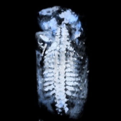
It is a great advantage to diagnose fetal skeletal dysplasia with 3D/4D technology because bone mineralization can be assessed and it is possible to compare bones (e.g., the spine with the skull bone and lavage bone), according to Dr. Peter Conner, PhD, of Karolinska University Hospital in Sweden. With 4D sequences, the fetal movement pattern and soft tissues can be assessed and, hence, malalignments can be diagnosed. Furthermore, with 4D it is possible to see fractures in both the lavage bone and ribs by using skeletal view visualization. In this mode, the skeletal structures are enhanced and soft tissues are suppressed.
A combination of better usage of 3D technology and knowledge of 3D technology in skeletal dysplasia will make it possible to correctly assess and diagnose many skeletal dysplasias in the first trimester. This is a major improvement, as most skeletal dysplasias are not currently diagnosed until the second or third trimester in families without a known family history of the condition, according to Conner. Adaptive volume enhancement technology, which suppresses unwanted signal such as noise/speckle, could also potentially facilitate the early diagnosis of skeletal dysplasias.
 Fetal spine evaluated using skeletal view rendering with a sepia color map (left) and standard color map (right).
In fetal brain examinations, 3D imaging provides a full volume of the fetal brain. Adaptive volume enhancement and true-to-data rendering -- which combines adaptive volume enhancement, scan conversion, and rendering to yield high resolution and fewer image approximations -- can also improve clinical value and make it easier to perform the study. 3D ultrasound fetal brain measurements are more accurate and reproducible than their 2D counterparts, according to Selbing.
 Fetal brain on an A-plane image before volume enhancement (left) and after adaptive volume enhancement was applied to the raw image volume (right).
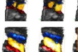
Furthermore, in fetal cardiology, 4D technology with color Doppler and a transparent image view can provide an improved view of the spatial relationship of the cardiac anatomy, specifically the relationship between both ventricles, the greater vessels, and the cardiac chambers, said Dr. Marius Vicea Calomfirescu of Bucharest, Romania, in a talk at ISUOG 2015.
Making the most of 3D/4D
When relying on 2D ultrasound scans, diagnosticians may completely miss the anatomical structure or feature they're looking for. There's a different risk with 3D ultrasound images; the anomalous structure is more likely to be acquired but may be missed by either the software or the diagnostician. Although image analysis software is making impressive strides toward significantly improved automated diagnosis, skilled technicians and diagnosticians are still required to produce a proper diagnosis in a reasonable amount of time.
Adaptive processing algorithms can be used to improve the image quality of the 3D ultrasound image volumes, helping to avoid misses of any structures. 3D/4D volumes consist of data in arbitrary planes, so it's important to perform adaptive volume enhancement in all dimensions. This ensures that structures unavailable in the 2D multiplanar reformatting (MPR) plane are not disregarded, that important diagnostic information is not discarded, and that artifacts are not introduced in the volume.
Diagnosis is often made during the scanning procedure, so it's essential that volume processing be performed in real-time. Adaptive algorithms will analyze every voxel to distinguish true information from artifacts such as noise and speckle, and then enhance this true information for more precise visualization of lines and complex structures.
The improvements in 3D image quality brought about by volume enhancement and specific visualization techniques make ultrasound an easy choice for many operators. With true volume enhancement and true-to-data rendering, you will improve the ability to find important structures and potentially improve your diagnosis. These techniques allow you to get a near-real-time look at specific areas and make it easier than ever to get better pictures.
Isabelle Wegmann Hachette is a senior application manager for ultrasound at image enhancement software developer ContextVision. The comments and observations expressed herein do not necessarily reflect the opinions of AuntMinnie.com.