Three-dimensional diffusion-tensor MRI (DTI-MRI) examinations of the cervix may have discovered at least one reason why some pregnant women miscarry or go into labor prematurely, according to a U.K. study published online on 11 December by the British Journal of Obstetrics and Gynaecology.
Researchers from the University of Leeds are using 3D DTI-MRI to view fibers in the cervix that help maintain its structure and prevent a fetus from descending into the birth canal before his or her due date.
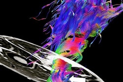
 The 3D MR image offers a horizontal view across the cervical canal and reveals encircling fibers, which give support to the fetus and prevent it from entering the birth canal too soon. Image courtesy of the University of Leeds.
Approximately 25% of miscarriages that occur during the fourth to sixth month of a pregnancy happen because of a weakened cervix. High-resolution 3D DTI-MR images thus far have provided details of the microstructure of the organ, which could explain why certain women have pregnancy issues, said lead author James Nott, an anatomy demonstrator and doctoral candidate at the university's Faculty of Medicine and Health.
"By applying the imaging techniques that have been used on the brain, we can get a much clearer understanding of the tissue architecture that gives the cervix its unique biomechanical properties," he said.
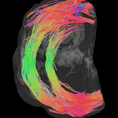
In this study, 3D DTI-MR images evaluated cervical tissue based on water content to discover a fibrous structure that extends along the upper part of the cervix, with fibers that are much more visible at the point where the cervix attaches to the womb.
During pregnancy, the fibers provide a strong supporting barrier that keeps the fetus and amniotic sac in place to preventing micro-organisms from entering the uterus. MRI scans show these supporting fibers are less prominent further down the cervix as it joins the birth canal.
During labor, the body releases chemicals that result in the cervix opening, allowing the baby to enter the birth canal. However, medical conditions that can occur early in a pregnancy could result in a failure of the cervix to support the baby and lead to a miscarriage or premature birth.
"This study's findings have encouraged us to explore new imaging techniques to check the integrity of these fibers before or during pregnancy in order to identify at-risk mums, intervene earlier, and so prevent late pregnancy loss and preterm birth," said study co-author Dr. Nigel Simpson, associate professor in obstetrics and gynecology at the university.
The study was funded by Cerebra, a charity for children with brain conditions.