Football's World Cup is 10 months away, but already the medical community in Russia is starting to warm up for the event. Researchers have unveiled the findings of a significant study of groin injuries in professional football players from Russian Premier League side FC Locomotiv Moscow.
"Given the complicated nature of groin pain diagnosis, MRI is an essential tool for accurately determining the cause of patients' complaints, applying equally to the acute condition or repetitive trauma in players with overuse syndrome," noted lead author Dr. Valentin Sinitsyn, professor and head of radiology at the Federal Center for Rehabilitation and Treatment in Moscow, in an e-poster presentation at the 2017 meeting of the European Society of Musculoskeletal Radiology (ESSR).
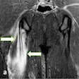
 Coronal T2-weighted fat-suppressed MR image shows a partial tear of pectineus muscle in a 27-year-old football player who presented with hyperintense hemorrhage and tearing up to 50% of muscle fibers (green arrow); focal defect shows partial retraction of muscle fibers. Grade 2 strain (moderate partial muscle tear) was positive for significant fiber disruption, probably including some retraction, fascial injury, and intermuscular hematoma. All images courtesy of Dr. Valentin Sinitsyn; Dr. Maria Lisitskaya, PhD; and Dr. Sergey Khaykin, PhD, presented at the 2017 annual meeting of the ESSR.
The authors studied MRI data from 28 players who complained of groin pain to their team doctor and were referred for imaging between July 2014 and May 2016. The mean age of the cohort was 29.7 (range 25 to 34 years old). One of the co-authors, Dr. Sergey Khaykin, PhD, is a sports physician at FC Lokomotiv.
Groin pain is typically an overuse injury due to excessive athletic activity. The term is the preferred label that refers to a spectrum of musculoskeletal injuries that occur in and around the pubic symphysis, and that share similar mechanisms of injury and common clinical manifestations, they pointed out.
MRI scans were performed in Sinitsyn's department on three machines: 1.5-tesla Avanto from Siemens Healthineers, 1.5-tesla Optima MR 450 from GE Healthcare, and 3-tesla Discovery MR 750 from GE. The standard protocol included a large field-of-view (36 to 40 cm) coronal T1-weighted spin-echo, coronal short time inversion recovery (STIR), and axial and coronal T2-weighted or proton density-weighted fat-suppressed imaging of the pelvis.
In players with muscle strains, sagittal proton-density-weighted fat-suppressed imaging of the affected hip was performed, in addition to coronal and axial T2-weighted or proton-density weighted fat-suppressed imaging. Also, the researchers acquired a fluid-sensitive T2-weighted fat-suppressed image in the coronal and axial plane to detect bone marrow edema to check for the presence of osteitis pubis. To help identify inguinal hernia, they obtained real-time functional imaging of abdominal wall motion.
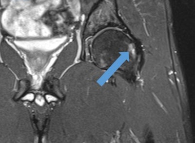 Coronal T2-weighted fat-suppressed MR image of the hip shows cam-type femoroacetabular impingement in a player. Note the increased signal intensity of subchondral femur head due to edema and cystic changes (blue arrow).
"MRI scans were reviewed for bone marrow edema in and around the pubic symphysis. Any abdominal wall, inguinal, femoral, or internal hernia was recorded," the authors wrote. "Tendinous injury involving the rectus abdominis and the adductor tendons (pectineus, adductor longus, adductor brevis, adductor gracilis, and adductor magnus) were reviewed. Tendon findings were classified as pathologic when there was a tendon enlargement, peritendinous fluid with or without intratendinous fluid signal intensity, partial tear, and complete disruption."
What were the key findings?
They divided all cases of pubalgia into two categories: acute traumatic pubalgia and exacerbation of pain caused by chronic conditions.
The first group (39% of patients) consisted of traumatic lesions of muscles and tendons. Three players had structural muscle injuries with partial tear of muscles and their tendons and intramuscular hematomas, two players had a complete muscle tear with retraction, and six players showed no visible signs of tissue damage and presented with mild edema. Three players had acute hip joint trauma consisting of capsule injury or acetabular labral tear.
The second group (61%) included players with groin pain as a manifestation of overuse syndrome induced by repetitive microtrauma. Hip joint related disorders were seen in two players, and also there were spine-related conditions in two players.
In seven players with suspected inguinal hernia, MRI showed spermatic cord edema. True inguinal hernias were seen in two players, three players had an occult hernia, and two more had mesh-related edema after laparoscopic repair surgery. Six players had signs of osteitis pubis on MRI.
"The anatomy of the pubic symphyseal region includes a number of interrelated muscle attachments that are located in close proximity to one another. The interrelation of these muscle attachments causes complex interactions between the forces exerted through the muscles across the pubic symphysis," Sinitsyn and colleagues explained to ESSR members at the annual meeting held in mid-June in Bari, Italy.
 Inguinal hernias, whether occult or obvious, and lipomas of the spermatic cord or round ligament are important etiologies to consider in the diagnosis of groin pain. This axial T2-weighted MR image of the pelvis shows a spermatic cord lipoma.
Tendon injuries are often overuse injuries or a result of a single traumatic event, they continued. Athletic muscle injuries present as a heterogeneous group of muscle disorders. The most widely used classification is an MRI-based graduation defining four grades: grade 0 with no pathological findings, grade 1 with a muscle edema only but without tissue damage, grade 2 as partial muscle tear, and grade 3 with a complete muscle tear.
Inguinal hernias, whether occult or obvious, and lipomas of the spermatic cord or round ligament are important etiologies to consider in the diagnosis of groin pain, the authors wrote. Many patients do not complain of a bulge, but instead complain chiefly of groin pain, unaware of the vast differential diagnosis list involved.
These hernias are often diagnosed with history and physical exam alone, and treated accordingly. An occult hernia can be a cord lipoma or indirect hernia sac that tracks along the spermatic cord within the inguinal canal creating compression on the ilioinguinal or genitofemoral nerves, they added. Lipomas of the cord and round ligament cause similar pain to that of a hernia.
Lower abdominal and pelvic pain
Osteitis pubis is a noninfectious inflammatory process of the pubic symphysis commonly seen in football players, and previous trauma, overuse, and vaginal delivery are all risk factors, they told ESSR delegates. Because of chronic repetitive shear, distraction injuries, and unbalanced tensile stress from the muscle attachments of the pubic symphysis, it is thought to result from instability of the pubic symphysis.
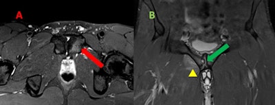 Mild osteitis pubis in a man. A: Transversal T2-weighted fat-suppressed MR image reveals parasymphyseal hyperintensity extending from the subchondral plate of left pubic body, which reflects edema due to increased stress response and areas of trabecular microtrauma. B: Coronal T2-weighted fat-suppressed MR image shows articular surface irregularity, erosions, osteophytes, and subchondral cyst (green arrow). In addition, there is an edema of myotendinous attachments of adductor seen as a coexistent tendinopathy and grade 1 strain (yellow arrowhead).
"As radiographic technology has improved, osteitis pubis is now recognized as a cluster of different injuries to the muscles, tendons, and osseous structures of the lower abdominal wall and pelvis," the researchers pointed out. "The mechanism of injury in athletic pubalgia combines two physical phenomena: repetitive motion injury and muscle development asymmetry."
Substantial amounts of bone marrow edema at the pubic symphysis can occur in asymptomatic football players, and it is only weakly related to the development of osteitis pubis. Therefore, MRI should be used to confirm the clinical suspicions provided by the patient history and a physical examination, they noted.
Injuries under the microscope
Athletic pubalgia (sports hernia) is a cluster of distinct injuries that are grouped together because of the common location of pain, overlapping activity triggers, and lack of physical exam findings. Most injuries to athletes result from a single action or collision, according to Sinitsyn and colleagues.
"Although groin injuries may be acute, they more often have an insidious onset and progress over a period of weeks or months. Patients may experience alternating episodic exacerbations and periods of improvement, or they may have gradually progressive symptoms," they wrote.
The differential diagnosis of groin pain in athletes is complicated by the fact that various pathologic entities may cause similar clinical signs and symptoms. Moreover, there are often overlapping findings at physical examination. The pathophysiologic conditions that cause groin pain are complicated and poorly understood.
Looking ahead to the 2018 FIFA World Cup, unfortunately for staff at the Federal Center for Rehabilitation and Treatment, another large city hospital has been appointed as the main medical facility for the event, which runs from 14 June to 15 July and will include 32 teams, Sinitsyn told AuntMinnieEurope.com. "But we do hope that we also will be involved because of our experience with sports imaging."
For free access to the authors' full range of clinical cases and images presented at ESSR 2017 and available on the European Society of Radiology's EPOS database, click here.


