MANCHESTER - In a new study presented by experienced investigators at this week's U.K. Radiological Congress (UKRC), well-trained reporting radiographers achieved high levels of accuracy and boosted cost-effectiveness in the interpretation of CT head scans. The findings look set to increase interest in role extension. The findings look set to increase interest in role extension.
Head injuries are becoming increasingly common, noted Paul Lockwood, senior lecturer at the School of Allied Health Professions, Canterbury Christ Church University, in the U.K. The Acquired Brain Injury Forum estimates the number of patients with head injuries attending U.K. district general hospitals is more than 350,000, or around 558 per 100,000 of the population each year, and this represents a 33.5% increase in the last 10 years of admissions for severe traumatic brain injuries.
Along with Keith Piper, PhD, principal lecturer in radiography at the same institution, Lockwood conducted a preliminary study to assess the diagnostic performance of a small group of reporting radiographers and senior radiologists in a clinical practice undertaking CT head interpretation. They applied a multiple reader, multiple case, alternative free response receiver operating characteristic (ROC) methodology, and used an image bank of 30 CT head examinations, with a 1:1 ratio of normal to abnormal cases.
The authors established a reference standard by double reporting the original reports using two additional independent consultant radiologists with arbitration of discordance by the researchers. Eight observers from six National Health Service (NHS) hospital groups in the south of England participated in the study. The group compared the results for accuracy, agreement, sensitivity, and specificity.
The six radiographers achieved a mean sensitivity of 88.7%, specificity of 95.6%, and accuracy of 92.2%, while the comparable figures for the radiologists were 83.4%, 90%, and 86.7%.
In related research, the same lead author carried out an audit of six radiographers who completed an accredited academic program in clinical reporting of CT head exams. For 3,008 reports, their mean accuracy score was 90.7%, and they recorded a mean sensitivity of 86.9% and specificity of 94%. The most common errors were associated with herniation, lacunar infarctions, and subtle fractures (false negatives), as well as involutional changes, subtle infarctions, and ventricular dilation (false positives).
"The results suggest appropriately trained radiographers can successfully undertake to report CT head examinations to a high standard," Lockwood stated. "The adoption of both academic and clinical workload image banks that reflect disease examples and the prevalence that may logically be encountered in practice offers the potential for an accurate measure of performance of radiographers' abilities."
Furthermore, an economic evaluation in Canterbury confirms that introducing a skills mix reporting service model can benefit service delivery and bring a potential annual cost saving for a hospital of between 125,000 and 300,000 pounds (around 142,000 and 340,000 euros).
"Research into recorded discrepancy/error audit data for potential detrimental risk to patient outcomes identified a paucity of evidence, and recommends further research is needed," Lockwood concluded.
With its inception in 1994, Canterbury Christ Church University became the first facility in the world to offer an accredited program for radiographers.
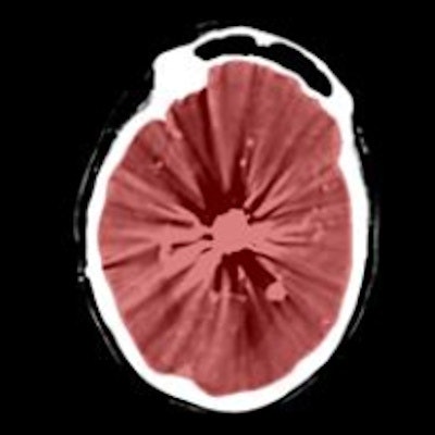
Editor's Note: The image on the home page shows an axial CT view of final cranial cavity segmentation for a patient with a large metallic foreign object between cerebral soft tissue. Image courtesy of Ajay Patel from Radboud University Medical Center in the Netherlands.