In a study that could inform clinical trials of multimodality imaging in radiotherapy, researchers in Germany and Slovenia have investigated correlations between dynamic PET and MRI in individuals with head and neck cancer.
Multimodality PET/MRI is gaining increasing attention in radiation oncology following the arrival of commercial hybrid scanners in hospitals in 2011. Researchers hypothesize that the more comprehensive characterization of the tumor microenvironment that such scanners promise could enable more effective treatments, tailored to the biology of individual tumors. This could include the use of radiosensitizers and dose painting in patients with radioresistant tumors. Multimodality imaging could also assist in monitoring patients' responses to treatments targeting hypoxia and tumor angiogenesis (Medical Physics, 20 April 2017).
Detecting tumor hypoxia
Static PET using fluorine-18-labeled fluoromisonidazole (FMISO) can detect tumor hypoxia, an indicator of radioresistance. Dynamic FMISO-PET, on the other hand, provides richer information on the perfusion and retention of the tracer by the tumor and is a more reliable test for hypoxia. However, protocols for dynamic FMISO-PET are complex and consequently rarely used, including in research.
Dynamic contrast-enhanced MRI (DCE-MRI) characterizes the perfusion and permeability of tumor vasculature. It is a potential alternative to dynamic FMISO-PET, motivating researchers from University Hospital Tübingen and the University of Ljubljana to investigate correlations between the PET- and MRI-based dynamic techniques.
"Optimization of a FMISO-PET/DCE-MR multimodality imaging protocol for cancer patients would be much easier if the relations between FMISO-PET and DCE-MRI kinetic parameters were known," wrote first author Urban Simoncic and co-authors in the paper.
3-tesla scans
The researchers imaged six individuals with head and neck cancer with separate FMISO-PET and DCE-MRI scans on the same day, before the start of a course of salvage radiotherapy. Several dynamic series of FMISO PET frames were acquired with the patient positioned as if for treatment, including immobilization using a mask. Static PET/CT images were also acquired two and four hours following injection. The final acquisition was used to generate maps of tumor-muscle ratio (TMR), a measure of tracer uptake by the tumor that was included in the correlation analysis.
DCE-MRI data was acquired on a 3-tesla scanner with a time-resolved angiography with interleaved stochastic trajectories (TWIST) sequence and a standard head and neck coil, without treatment immobilization. FMISO-PET and DCE-MRI datasets were coregistered using CT data from the static PET/CT and anatomical T2-weighted MR images acquired in the same session as the DCE-MRI data.
The kinetic behaviors of FMISO and the gadolinium-based contrast agent were analyzed in individual voxels using two separate two-tissue compartment models, each with its own set of kinetic parameters. Correlations between the modalities using paired parameters were quantified using Pearson's correlation coefficients.
The tumors ranged from nonhypoxic to severely hypoxic, according to tumor uptake in the TMR maps. High correlation was revealed between vp and Vb, the respective vascular fractions in tissue for the two modalities, with a median correlation coefficient of 0.71. Values ranged from 0.69 to 0.74 in four of the six, while two demonstrated moderate correlations with values of 0.42 and 0.46.
Correlations between other model parameter pairs, which included transport rate constants for both modalities and a measure of the FMISO uptake rate into tissue, were lower overall and varied substantially between patients. The authors concluded that the parameters were unrelated and that further investigations of the physiological and physical processes responsible for the low correlations are needed.
"Observed correlations ... indicate that DCE-MR imaging provides similar information as the kinetic analysis of dynamic FMISO-PET data," the authors wrote. "However, the information from both imaging modalities is not exactly the same. Therefore, FMISO-PET/DCE-MR multimodality imaging might be a good choice if the dynamic FMISO-PET imaging is not possible."
Jude Dineley is a freelance science writer and former medical physicist based in Bavaria, Germany.
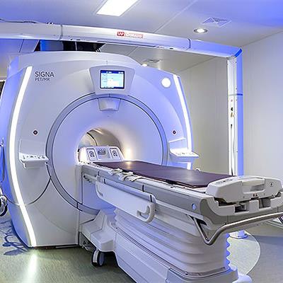
Editor's note: Image of PET/MRI system on home page courtesy of Mikael Stiernstedt.
© IOP Publishing Limited. Republished with permission from medicalphysicsweb, a community website covering fundamental research and emerging technologies in medical imaging and radiation therapy.