PET is widely employed in the clinic to track molecules within the body with high sensitivity. Currently, the vast majority of PET scans are performed using the radiotracer F-18 FDG to image tumors. But according to Charalampos (Harry) Tsoumpas, PhD, a lecturer in medical imaging at the University of Leeds, PET has a lot more to offer when it comes to clinical applications.
"When PET was combined with CT, that was a big breakthrough that changed clinical practice," Tsoumpas said. "But I believe that molecular imaging is just starting."
 Harry Tsoumpas with Dimitra Darambara, chair of the IOP Medical Physics Group.
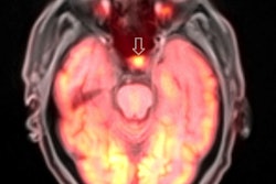
Speaking at the IOP Medical Physics Group's recent meeting, "Up and Coming Techniques in Medical Physics Translated into Clinical Practice," Tsoumpas explained there are many other potential radiotracers that could be employed, perhaps with the ultimate goal of developing an individual tracer for every disease. PET can also move beyond the imaging of cancer, for example, enabling visualization of ruptured or high-risk coronary plaque using F-18 fluoride tracers.
Another area with great potential is tracking response to therapy. Performing weekly PET scans after therapy, for example, could help physicians personalize a patient's treatment regime. "It is very difficult to design a therapy that will work for everybody," Tsoumpas said. "With PET, we can see how the body responds before any morphological changes occur. This provides insight to change therapy where needed."
Software advances
With these emerging applications in mind, how can physics help progress clinical PET? Firstly, Tsoumpas explained, new developments in mathematical and computational modeling have advanced PET image reconstruction software and techniques. For example, regularized image reconstruction, which incorporates prior knowledge about the image, provides more accurate PET quantification. This approach has reached the clinic, he noted, but is not used routinely, with much research work still needed.
The physical modeling of factors such as attenuation and scatter, randoms, and detector response are now included in clinical scanners. But other challenges remain, such correction for positron range (the distance traveled by positrons before annihilation), which is not yet offered in commercial software and needs further investigation. This effect can introduce inaccuracy into PET quantification and is particularly problematic for gallium-68-based radiotracers that emit highly energetic positrons.
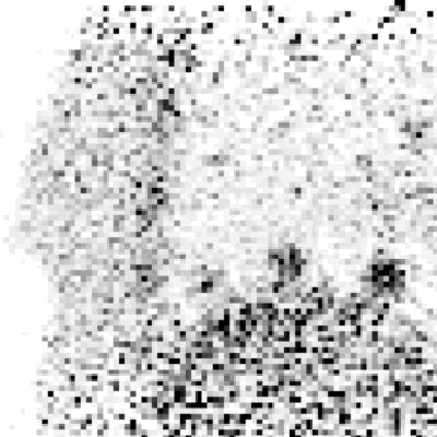
 The animated images show the visual improvement achieved by including regularization within motion-compensated PET image reconstruction, for three lesions in a computational phantom. Original source: C Tsoumpas, Physics in Medicine and Biology, Vol. 58:6, pp. 1759-1773.
Physiological modeling of tracer kinetics can also help to improve the accuracy of image reconstruction, Tsoumpas said, highlighting the development of dynamic whole-body 4D reconstruction. This technique, which is currently being translated into the clinic, uses multiple bed positions and snapshots over time to measure the time-dependent spatial distribution of tracers in blood and tissues.
Another active area of research is motion correction. Respiratory motion, for example, creates movement in the range of 6 mm to 18 mm, and can cause image blurring, limit tumor detectability, and result in underestimation of tracer uptake values. "We need to find ways to measure respiratory motion accurately," Tsoumpas said. "PET will have increased diagnostic value with respiratory motion correction."
He noted that several consortia have been set up to solve such problems. In the U.K., for example, STIR (software for tomographic image reconstruction) provides an open source library of software for use in tomographic imaging, with a current emphasis on (iterative) image reconstruction in PET and SPECT. More recently, the CCP PET-MR (collaborative computational project in PET and MRI) was established to bring together PET and MR image reconstruction expertise to develop a PET/MR software infrastructure.
Hardware happenings
Despite being a software researcher, Tsoumpas admitted that recent hardware developments have proved even more exciting. He cited the introduction of integrated PET/MRI scanners, noting there are about 100 such systems currently in clinics worldwide. "PET/MRI is a fantastic tool," he said. "It offers a lot of scope for further research."
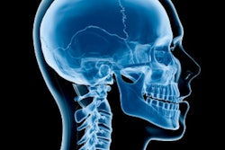
PET/MRI can play a key role in early detection of dementia, for example. And the ability to combine different tracers and different MR sequences opens up possibilities way beyond simply adding two imaging modalities together.
There are, however, many unmet challenges for PET/MRI, not least the need to identify and further investigate principal clinical applications. The acquisition time for PET/MRI scans is also longer than needed for PET/CT -- approximately 50 min as opposed to 20 min -- although this longer acquisition does provide a higher signal and may enable tracking of kinetics. Finally, there's the challenge of how to perform attenuation correction without access to electron density data provided by CT.
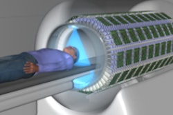
Another recent hardware breakthrough is the EXPLORER total-body PET scanner, under development by a research consortium led by UC Davis in the U.S. This 2 m-long scanner will image the entire body simultaneously, providing 40 times greater sensitivity. This gain could be used to lower the radiation dose by a factor of 40, enabling longitudinal imaging of tracers over several weeks and turning PET into a "near-radiation-free" screening tool. Alternatively, the sensitivity gain could enable imaging in just a few seconds, enabling visualization of nuclides with short half-lives.
The UC Davis team was awarded $15.5 million U.S. (14.6 million euros) from the National Institutes of Health to build this world-first total-body PET scanner. They have already created a prototype for medium-sized-animal (for example, canine) imaging and a mock clinical system, with a clinical device slated for next year.
Finally, Tsoumpas cited the introduction of time-of-flight (TOF) capability on PET systems. TOF provides more accurate localization of annihilation events in the patient and increases the image quality and tracer sensitivity. Current clinical TOF systems have a timing resolution of around 350 ps to 400 ps. But recent research shows the time-of-arrival of 511 keV photons at the detector can be identified with approximately 100 ps timing precision. Meanwhile, the research consortium COST Action FAST aims to make it even faster. TOF can also enable many new PET applications, such as the use of limited angle tomography and the creation of wearable PET systems.
"Physics developments may change the way that we perform imaging with radioactivity in the future," Tsoumpas concluded.
© IOP Publishing Limited. Republished with permission from medicalphysicsweb, a community website covering fundamental research and emerging technologies in medical imaging and radiation therapy.