Photoacoustic computed tomography (PACT) can provide noninvasive real-time imaging of significant anatomical and physiological information. The strong acoustic waves generated by the pulsed laser used for illumination deliver high image resolution and sensitive detection. However, the size, bulk, and cost of a pulsed laser, and the expensive and laborious procedures required to maintain it, have deterred clinical adoption of PACT.
A single xenon flash lamp may be a practical and viable alternative illumination source, according to researchers at the Optical Imaging Laboratory of Washington University in St Louis. They tested its capabilities with biologically mimicking phantoms, with ex vivo tissues and in vivo with a laboratory mouse. The resulting images were compared with those acquired from a pulsed-laser based PACT (Journal of Biomedical Optics, 24 October 2016, Vol. 22:4, pp. 041003).
Compared with other potential alternative illumination sources such as LEDs or pulsed laser diodes, xenon flash lamps have high pulse energy, microsecond pulse width, and a much wider spectrum. The research team used a xenon flash lamp (L4634 from Hamamatsu Photonics) with a borosilicate glass housing and a light spectrum spanning from 240 to 2000 nm. The illumination light covered an imaging area of about 20 cm2. The addition of two 0.1 µF capacitors enabled the flash lamp to operate at its highest possible output.
 Terence Wong from the Optical Imaging Laboratory at Washington University in St Louis.
Lead author Terence Wong and colleagues from the laboratory of Lihong Wang used a xenon flash lamp to image a phantom containing a 1.5 mm diameter latex cord configured in an "eight" shape, using four focused ultrasound transducers with various center frequencies. They determined that a transducer with a center frequency of 0.5 MHz generated the highest contrast-to-noise ratio (CNR) and produced the best images with the xenon flash lamp. When comparing images produced by flash lamp excitation and the optimal transducer with those produced by laser excitation, similar features could be observed and the quality of both sets of images was comparable.
The researchers then acquired image sets from each illumination source of seven pencil leads placed on agar-water gel, a latex cord surrounded by chicken breast tissue, a blood-filled tube embedded in agar-water gel, and a mouse. The pencil leads were used to characterize the imaging resolution of the xenon flash-lamp-based PACT. They determined that the average in-plane imaging resolution within a 25-mm radius area was approximately 1.5 mm for both sources, and that the short pulsed xenon lamp preserved imaging resolution well.
To determine the maximum depth of a recognizable photoacoustic image and to simulate deep-tissue imaging, the researchers placed two crossed latex cords within slices of chicken breast ranging from 1.0 to 4.0 cm in thickness. Cross structures could be seen in all images to a maximum penetration depth of 3.5 cm, with a CNR of 2.9. Use of multiple flash lamps could improve the CNR and the penetration depth.
 Lihong Wang from the Optical Imaging Laboratory at Washington University in St Louis.
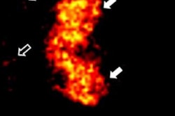
Imaging of blood was simulated using blood-filled tubes embedded 1 cm inside an agar water gel. To compensate for the fact that blood is less absorbing than a latex tube, the researchers used a ring-shaped ultrasonic transducer array with 512 elements to improve the signal-to-noise ratio via 1,000 times averaging. They also imaged the body, tail, and legs of a live mouse immersed in water with 5,000 times averaging. The blood-filled tube could be observed with sufficient contrast and the different structures of the mouse could be seen.
These findings show that xenon flash-lamp-based PACT has the potential for use in many cost-effective biomedical and clinical applications. The team noted this illumination source, with its ability to acquire images at 3.5 cm tissue depth using a single lamp, would meet the requirements of many biomedical applications, such as imaging breast cancers.
"Using a single xenon flash lamp with minimal optical and acoustic components, a complete PACT system can be appealingly compact, portable, and inexpensive, facilitating the use of photoacoustic tomography in clinics and global healthcare," the authors wrote.
"One quickly apparent clinical application of a xenon flash lamp photoacoustic imaging system is imaging skin melanoma with accurate depth and volume," Wong told medicalphysicsweb. "Extracting these parameters in vivo could significantly improve melanoma patient management and avoid the need for a second or excisional biopsy."
© IOP Publishing Limited. Republished with permission from medicalphysicsweb, a community website covering fundamental research and emerging technologies in medical imaging and radiation therapy.