Interventional radiology is well-established in clinical practice of human medicine, but to date it has had very limited application in marine animals in an aquarium setting. The development of new techniques, however, could increase the rate of reproductive success in cetaceans, particularly in endangered species for which conservation is a priority. Interventional radiology techniques could have a vital role to play in bolstering them.
Through the cooperation of veterinary specialists and radiologists, it was possible to minimize the time required to cross the cervix with the endoscope in a beluga whale and provide, for the first time, the opportunity to place an intrauterine catheter inside the uterus of a bottlenose dolphin. This procedure eliminated the need to cross the dolphin's cervix with the whole endoscope.
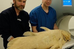
 Combined endoscopic-interventional radiology approach for beluga whale fertilization.
The whale underwent moderate sedation for the procedure. A 0.035-inch Jagwire angled stiff hydrophilic guide wire (Boston Scientific) was inserted for endoscopy guidance to navigate through the lumen of the narrow cervix into the uterus, reducing the total time required for insemination. Once the endoscope was in, the standard technique was completed by the subsequent advancement through the working channel of a 6-French simple catheter in the uterine cavity for semen inoculation.
A similar approach was then tested in two bottlenose dolphins. In the dolphins, only an intrauterine catheter was passed through the cervix instead of the whole endoscope and guide wire. This minimized the necessity to insufflate the uterus with air, which has the potential to affect postinsemination uterine motility. Once the catheter is placed in the uterus, transabdominal ultrasound may be of value in smaller cetaceans to track further catheter advancement.
Artificial insemination
The use of artificial insemination techniques has notably increased the opportunities for cetacean reproduction and population management in recent years. However, due to the singular reproductive anatomy of the genital tract of female cetaceans, insemination techniques are complex and time-consuming, requiring very specific endoscopic equipment and experienced professionals to successfully manage intrauterine insemination.
 Longitudinal section of the reproductive female porpoise (Meek A. The reproductive organs of cetacean. J Anat. 1918;52:pt 2, pp.186-210).
The most common technique requires endoscopic navigation through the female genital tract, releasing frozen-thawed semen in the uterine horns as close as possible to the fallopian tubes. Intravaginal insemination is significantly less effective, requiring larger dosages of semen and higher sperm quality to obtain similar success ratios. The goal, therefore, is to deposit semen as far as possible into the uterine tract to minimize the volume and concentration of semen required, optimizing the use of the valuable stored samples.
The complex cetacean genital anatomy combined with a small colon and rectum precludes the possibility of direct rectocervical approach, whereby the catheter is inserted into the vagina, and rectal manipulation facilitates the passage of the insemination pipette into the cervix or distal uterine body as is performed in most domestic species.
In general, cetaceans have vaginal folds associated with cervical structures that vary in complexity between species. The function of this vaginal/cervical complex is unknown, but it has been hypothesized it forms a seal that prevents saltwater contamination of the ejaculate and/or acts as a selective mechanism in polygamous mating systems.
Traditionally, visual guidance during insemination has relied on endoscopic advancement through the vaginal folds and cervix before entering the uterus. Until now, professionals needed to guide the entire flexible endoscope through the whole genital tract while insufflating air before releasing the semen with a very long custom-made catheter through the working channel.
This interventional approach might improve uterine motility postinsemination, reduce the possibility of uterine contamination, and increase conception rates. In addition, the possibility of uterine catheterization can in fact open possibilities for more advanced assisted reproductive techniques, such as embryo collection through the application of uterine lavage within a closed system, which was never achieved before in cetaceans, but has already been carried out by this group in the previously mentioned bottlenose dolphins.
Dr. Raúl García is a radiologist at Biomedical Imaging Research Group (GIBI230) at La Fe Polytechnics and University Hospital and La Fe Health Research Institute in Valencia, Spain. He is currently active in all aspects of interventional radiology, but is particularly involved with interventional oncology and interventional pain management. He collaborates with Oceanografic Aquarium in Valencia to perform minimally invasive procedures in marine mammals.
Todd Robeck, DVM, PhD, is the vice president of theriogenology at SeaWorld Parks and Entertainment. He is responsible for the development and application of assisted reproductive technologies to the animal collection at SeaWorld Parks and Entertainment. He has been the principle or co-principal investigator in developing the first successful methodology for artificial insemination in the killer whale, bottlenose dolphin, Pacific white-sided dolphin, the beluga whale, and the Magellanic penguin.
Daniel García-Párraga, DVM, is a specialist in veterinary medicine and the director of animal health research and conservation at Oceanogràfic, Veterinary Services, Ciudad de las Artes y las Ciencias in Valencia, Spain.
Dr. Luis Martí-Bonmatí, PhD, is the director of medical imaging at La Fe University and Polytechnic University Hospital, and chief of radiology at Quirón Hospital in Valencia, Spain. He is also professor of radiology at Valencia University.
Gisele Montano, DVM, is a research scientist at SeaWorld & Busch Gardens Reproductive Research Center (SWBGRRC) in San Diego. She collaborates on various projects on dolphins, beluga, killer whales, and other species of cetacean, as well as terrestrial animals, including penguins, elephants, and rhinos. She is also a consultant for Dolphin Adventures in Mexico on the reproductive management of their bottlenose dolphin population.
The comments and observations expressed herein do not necessarily reflect the opinions of AuntMinnieEurope.com, nor should they be construed as an endorsement or admonishment of any particular vendor, analyst, industry consultant, or consulting group.