A new MRI-based assessment approach for acute musculotendinous groin injuries in athletes shows good intra- and interrater reproducibility and may help improve clinician's confidence in MRI scoring of these injuries, a 6 July study in European Radiology found.
MR images were collected as part of a large, prospective study on athletes with acute groin pain. All athletes underwent MRI according to a standardized groin-centered protocol, and a system was developed to assess certain characteristics. The researchers, led by Dr. Andreas Serner from Aspetar orthopedic and sports medicine hospital in Doha, Qatar, and Dr. Frank Roemer from the radiology department at University of Erlangen-Nuremberg in Erlangen, Germany, found their standardized MRI assessment of acute groin injuries showed good reproducibility.
"Our approach may help improve clinicians' confidence in MRI scoring of acute groin injuries when comparing clinical findings to the parameters included in our study, such as injury location and severity," they wrote. "Furthermore the results of our study provide a strong foundation for further validation regarding prognostic MRI variables, which can determine the most relevant parameters to include in the reporting."
This can potentially simplify and substantially shorten time needed for assessment when applying this scoring protocol, the authors noted.
Why MRI?
Groin injuries are common and frequently occur in high-intensity team sports, such as football, accounting for up to 19% of all injuries, according to the researchers. To date, it has been unknown whether MRI data for acute groin injuries -- taking into account anatomical location, severity, and extent of injury, as well as associated imaging findings -- have adequate reproducibility.
A reliable MRI protocol and assessment system have been developed for long-standing groin pain, but an assessment system for acute groin injuries has been lacking -- until now.
Serner, Roemer, and colleagues developed a detailed multidimensional MRI assessment approach for acute groin injuries that focuses on musculotendinous injuries, including location, grading, and lesion extent, as well as other nonacute findings, and determines intra- and interrater reproducibility of such assessments.
The researchers collected MR images from a 1.5-tesla system (Magnetom Espree, Siemens Healthcare) as part of a large, prospective study on athletes with acute groin pain. A sports medicine physician recorded standardized injury history and clinical exam findings for the athletes. The study included 75 male athletes from 18 to 40 years old who were participating in competitive individual or team sports and presented at the specialized sports medicine hospital in Doha, Qatar, within seven days of acute onset of sports-related groin pain.
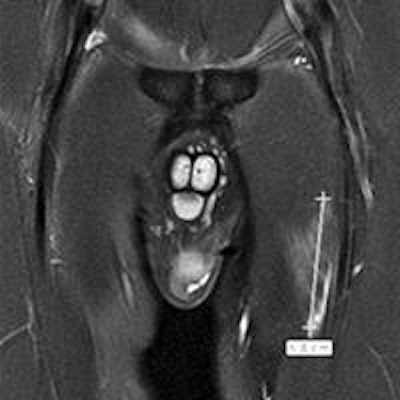
 Continuous measurements of edema in acute lesions. Above: Coronal proton density-weighted fat-suppressed image shows proximal distal extent of edema in an adductor longus lesion. Below: Corresponding axial proton density-weighted fat-suppressed image shows extent of edema in the anteroposterior and mediolateral directions. Bottom: Axial proton density-weighted fat-suppressed image shows a measure of the maximum thickness of perimuscular fluid-equivalent signal (double-headed arrow). All images courtesy of European Radiology.
MRIs were scored by two musculoskeletal (MSK) radiologists with 13 and 16 years of experience in applying semiquantitative scoring of MSK MRI in a research setting.
For intrareader reproducibility, one radiologist reassessed the MR images after an interval of six weeks to avoid recognition bias.
The protocol
The protocol the researchers developed assessed grade, location, and extent of muscle strains; perilesional hematoma; and other nonacute findings commonly associated with groin pain, such as location and extent of edema.
The researchers evaluated the following muscles in the groin region:
- Adductor group: the adductor longus, adductor brevis, pectineus, adductor magnus, gracilis, and obturator externus
- Iliopsoas (with iliacus and psoas separately when possible)
- Rectus femoris
- Obturator internus
- Rectus abdominis
- Sartorius
- Vastus medialis
- Tensor fasciae latae
Musculotendinous lesions were graded identically for acute and nonacute lesions on a scale from 0 to 3, with 0 being no imaging abnormality and 3 being complete musculotendinous disruption/tear or avulsion from the tendinous attachment.
In the 75 athletes, the researchers scored 85 different acute muscular lesions. One radiologist found 18 athletes were MRI-negative for acute lesion injuries, and the other radiologist found 17 athletes were MRI-negative for acute lesion injuries. The most commonly affected muscle was the adductor longus (42.7%), followed by the rectus femoris (16.3%).
The radiologists also scored 19 different nonacute lesions with the most common nonacute finding being pubic bone marrow edema, which was present in at least one side in 55% of the athletes. Other common pathologies were central disk protrusions/superior osteophytes and perisymphyseal sclerosis.
The researchers found almost perfect intra- and interrater agreement (κ = 0.81-1.00) for lesion presence/absence, number of acute lesions per athlete, and muscle location, as well as lesion grading and edema location in the axial plane. Edema location in the coronal plane had substantial agreement (κ = 0.70-0.85).
 Tendon waviness. Coronal proton density-weighted image shows tendon waviness (short, thick arrows) accompanying an extensive rectus femoris lesion.
Implications
"The almost perfect reproducibility for the presence and absence of acute lesions, as well as the specific muscle location, gives confidence that if a clinician is uncertain of a specific location of injury, the MRI assessment (when performed by an experienced radiologist) will provide a more detailed and decisive assessment," the researchers wrote.
However, as negative imaging results were reported in about 1 out of 5 acute groin injuries, the clinician would have to rely on the clinical exam findings in those cases.
"The groin region is anatomically complex, but using the current MRI protocol anatomic determination of specific muscles involved was possible," the study authors wrote. "Further research will have to show whether an abbreviated protocol may be applied to achieve comparable reliable scoring, consequently decreasing reading or assessment burden."