Using PET/CT as the reference standard, German researchers have found significant deficiencies with CT alone for the detection of bone marrow metastases in patients with malignant melanoma, according to a study published in the April issue of the European Journal of Radiology.
Contrast-enhanced CT's diagnostic accuracy was "limited," concluded the paper from Eberhard Karls University Tübingen, having missed a great majority of malignant lesions in the spine, pelvis, and thorax. CT also either missed or underestimated six of 11 cases with disseminated bone marrow involvement, which was defined as more than 10 bone marrow lesions or diffuse infiltration from the melanoma.
"Consequently, staging of melanoma patients using CT may lead to an underestimation of malignant bone and bone marrow infiltration with potentially fatal misjudgment of the prognosis and therapeutic options," wrote lead author Dr. Georg Bier and colleagues from the department of diagnostic and interventional radiology (EJR, April 2016, Vol. 85:4, pp. 732-738).
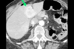
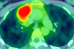
 A 59-year-old woman with disseminated bone marrow metastases of a malignant melanoma, which were mostly occult on CT images. Above: CT image. Below: PET image. All images courtesy of Dr. Georg Bier.
This inability of CT to detect the side effects of malignant melanoma would make it that much more difficult for oncologists to devise appropriate treatment for cancer patients.
PET/CT's track record
As Bier and colleagues noted, malignant melanoma is one of the most lethal types of cancer, and, once it metastasizes, patients face a poor prognosis.
While MRI and FDG-PET/CT provide high diagnostic accuracy in the initial staging of the disease, healthcare practitioners often turn to CT for follow-up exams to assess treatment response, because the modality is generally more accessible and is less expensive than MRI or PET/CT. But CT does have shortcomings.
"Whilst the sensitivity of CT is high for the detection of osteolytic bone lesions and can be enhanced by new image postprocessing methods, the detection of bone marrow involvement remains challenging, especially in patients with diffuse/disseminated bone marrow involvement/infiltration," the authors wrote.
So, the researchers sought to determine if CT on its own was up to the task for following up malignant melanoma patients. They specifically focused on disseminated and diffuse bone marrow metastases in patients suffering from malignant disease.
Bier and colleagues embarked on a retrospective, single-center study that enrolled 50 patients (27 male, 23 female) with a mean age of 61 (± 15.1 years) who had malignant melanoma stage IV cancer. All 50 subjects underwent whole-body FDG-PET/CT (Hi-Rez Biograph 16 or Biograph mCT, Siemens Healthcare) for initial diagnosis or follow-up between April 2008 and July 2015.
Two readers first interpreted the PET/CT scans, in which 18 patients (36%) were confirmed for metastases to the bone and/or bone marrow, while one patient had only bone metastases. The remaining six patients had no metastases and served as control group.
Once the PET/CT reference standard was set, the researchers then calculated sensitivity, specificity, and other aspects of the CT component of the scans alone. The presence of malignant or nonmalignant bone and bone-marrow lesions on contrast-enhanced CT was assessed by two radiologists, which included lead author Dr. Georg Bier, who recorded lesion location, character, and lesion size.
Lesion detection
With PET/CT, the two readers found a total of 594 bone lesions (11.5 ± 20.85 lesions per patient; range: 0-121 lesions) on PET/CT. That total included 495 (83%) malignant lesions or 9.02 (± 20.44) malignant lesions per patient. The remaining 99 lesions (17%) were classified as benign.
There were 197 (40%) malignant lesions located in the spine, followed by 113 malignancies (23%) in the pelvis, and 61 malignant lesions (12%) in the ribs. The humerus had 51 malignancies (10%) and the femur had 36 such tumors (7%).
Among the total were 385 medullary lesions (78%), which involved the inner regions of an organ or tissue.
Unfortunately for patients, CT most often missed medullary lesions. There were 300 (96%) medullary lesions that went undetected by CT. Those lesions had a mean size of 10 mm (± 6.2 mm, range of 3 mm to 63 mm). The modality only missed 13 (4%) of lytic lesions, which had a mean size of 5.0 mm (± 2.75 mm, range of 3 mm to 14 mm).
Most interestingly, given there were 197 malignant lesions in the spine, as seen on PET/CT, CT alone missed 153 malignant lesions (77%) in the spine. CT did not fare particularly well in the pelvis, missing 80 (71%) of the 113 malignant lesions in this area. (See chart.)
| CT lesion detection | |||
| Total malignant lesions | Undetected | Percent undetected | |
| Spine | 197 | 153 | 78% |
| Pelvis | 113 | 80 | 71% |
| Thoracic | 95 | 80 | 84% |
| Lumbar | 40 | 24 | 60% |
| Sacral | 33 | 20 | 61% |
| Cervical | 29 | 29 | 100% |
CT missed or underestimated six of 11 disseminated bone marrow involvement cases, compared with PET/CT, which provided accurate information on all 11 cases.
By not detecting melanoma malignancies, CT does a disservice to oncologists who need to determine the most appropriate treatment or upstage therapy for a particular patient's condition, according to the researchers.
"Moreover, a disseminated involvement of the bone marrow may induce a secondary bone marrow failure ... with the need for specific, immunoprotective therapy and blood component substitution," the authors noted.
In following up these patients, the researchers found that all 11 patients with disseminated bone marrow infiltration died within one year after their PET/CT scan, while six patients without disseminated bone marrow involvement died within the first year after PET/CT.
| Diagnostic accuracy of CT | |
| Lesion-based analysis | |
| Sensitivity | 37% |
| Specificity | 88% |
| Positive predictive value | 94% |
| Negative predictive value | 22% |
| Accuracy | 45% |
"PET/CT is a powerful tool to detect disseminated bone marrow infiltration and should be considered whenever clinical symptoms or blood sample results give suspicion of bone marrow involvement to detect disseminated bone marrow infiltration," the authors stated.
Bier and colleagues cited the study's small sample size and retrospective approach as limitations.
"Nevertheless, to our knowledge our study presents the first larger series of malignant melanoma patients with the rare occurrence of disseminated bone marrow involvement," they added.