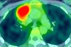
In the largest study of its kind to date, Austrian researchers have found that adding diffusion-weighted imaging (DWI) to FDG-PET/MRI can enhance the evaluation of patients with lymphoma, according to a study published online in Investigative Radiology.
 Dr. Chiara Giraudo from Medical University of Vienna.
FDG-PET/MRI with DWI achieved greater sensitivity and accuracy than FDG-PET/MRI and FDG-PET/CT and helped in the assessment of lymphomas with varying degrees of FDG avidity.
"We also can say that PET/MRI with DWI has an intrinsic lower radiation dose compared to PET/CT," said lead author Dr. Chiara Giraudo, from the Medical University of Vienna, who presented preliminary findings at RSNA 2015 in Chicago. "We strongly believe PET/MRI with DWI can represent the beginning of a new era for diagnosing lymphoma,"
Modality of choice
FDG-PET/CT has been considered for almost two decades as the modality of choice for both the initial staging and restaging of Hodgkin's lymphoma and the majority of non-Hodgkin's lymphomas.
The advent of FDG-PET/MRI has prompted studies to evaluate the hybrid modality's potential efficacy in this realm, although past studies have some shown some deficiencies with MRI for nodal staging, due in part to motion artifacts.
Still, early research has raised expectations of FDG-PET/MRI, because it can combine metabolic information from PET with MRI's high soft-tissue contrast resolution of anatomy. With the addition of DWI, the hybrid modality could also enhance the assessment of cell density.
Giraudo and colleagues also noted that DWI is "considered superior for some indolent non-Hodgkin's lymphoma subtypes," such as mucosa-associated lymphoid tissue (MALT), which are frequently not FDG-avid.
"The aim of our study was to assess the diagnostic value of PET/MRI including DWI and CT for Hodgkin's and non-Hodgkin's lymphoma," she said.
Imaging comparisons
Between January 2014 and April 2015, researchers recruited 19 males and 15 females with a mean age of 56 years who had histologically proven lymphoma and who were referred to a local tertiary care center for pretherapeutic staging or follow-up (Invest Radiol, 18 January 2016).
Among the patients, there were 20 subjects with indolent non-Hodgkin's lymphoma, 10 cases of aggressive non-Hodgkin's lymphoma, and four individuals with Hodgkin's lymphoma.
All scans were performed on the same day with a single injection of FDG to cover both procedures. The majority of subjects first received the FDG-PET/CT exam on a 64-detector-row hybrid PET/CT system (Biograph TruePoint 64, Siemens Healthcare) after five hours of fasting.
PET scans were performed 45 to 60 minutes after an intravenous administration of 300 MBq of FDG. Contrast-enhanced CT also was utilized to enhance the evaluation of extranodal disease and lymph nodes.
FDG-PET/MRI was performed directly after PET/CT with an integrated simultaneous hybrid PET/MRI device (Biograph mMR, Siemens) with a 3-tesla magnet. Total scanning time for entire PET/MRI procedure was approximately 120 minutes, including 15 to 20 minutes for the DWI sequence.
Researchers focused on 14 nodal regions, which included the lungs, liver, spleen, stomach, small and large intestines, both kidneys, bones, and soft tissue.
The study used identical criteria for all three imaging techniques when determining a positive result: at least one of the nodal or extranodal regions showed one or more focally increased tracer accumulations relative to surrounding tissues or mediastinal blood-pool activity for staging or to the liver for restaging.
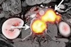
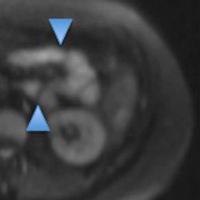
 A 39-year-old woman with a histologically verified MALT lymphoma of the small intestine. The color-coded, fused FDG-PET/CT (A) and FDG-PET/MRI (B) show no increased tracer uptake. However, the DWI image (C) and the corresponding ADC map (D) clearly demonstrate the extranodal lymphoma involvement (light blue arrowheads in C and D). Images courtesy of Dr. Chiara Giraudo.
Image comparisons
Among the 36 subjects were six patients who were scanned twice for staging and/or restaging, bringing the number of total exams to 40. Interestingly, the analysis of results concluded that all three imaging techniques discovered 28 positive lesions (70%) from the 40 exams.
But FDG-PET/MRI with DWI outpaced the other two modalities with no false-positive results, compared with five false positives (all non-FDG-avid mucosa-associated lymphoid tissue lymphoma) with FDG-PET/CT and four false positives with FDG-PET/MRI (see chart).
| Accuracy by modality for lymphoma | |||
| FDG-PET/CT | FDG-PET/MRI | FDG-PET/MRI with DWI | |
| Sensitivity | 82% | 86% | 100% |
| Specificity | 100% | 100% | 100% |
| Accuracy | 86% | 90% | 100% |
The region-based analysis found that FDG-PET/MRI with DWI detected a total of 113 nodal and extranodal positive regions, compared with 105 with FDG-PET/CT and 107 with FDG-PET/MRI.
Perhaps most important, FDG-PET/MRI with DWI correctly upstaged three patients with histologically confirmed lymphomas that were missed by FDG-PET/CT.
Giraudo noted the additional value of FDG-PET/CT with DWI is perhaps most evident in the fact that there were no false positives, compared with FDG-PET/CT, which missed the five non-FDG-avid MALT lymphomas.
"The additional value is for MALT lymphoma in our case," she said, "and we can expect the same positive results for other non-FDG-avid lymphomas, which are usually undetected or not so well-detected with PET/CT."
The Austrian researchers noted that despite having only 40 subjects, this study is the largest of its kind and the "first that also included a relevant number of lymphomas with variable FDG avidity, and the first that specifically evaluated the effect of including DWI in the PET/MR protocol -- clearly suggesting that FDG-PET/CT and FDG-PET/MR generally show a comparable performance in patients with lymphoma."
"Overall, we can conclude that PET/MRI with DWI can improve the diagnostic workflow of Hodgkin lymphomas," Giraudo said.