Wherever they come from, mummies intrigue us. They fascinate people of all ages, from each continent and of all sociocultural backgrounds. So it's only natural that they should also fascinate radiologists. At Toulouse University Hospital (CHU Toulouse), we were given the chance to study a mummy, discovered in Rennes, which was perfectly preserved.
A recent excavation (July 2012 to June 2013) of 8,000 m2 in Rennes city center undertaken by the French National Institute of Preventive Archaeological Research (Institut National de Recherches Archéologiques Préventives, INRAP), allowed the study of a Mendicant order convent: the Convent of the Jacobins.
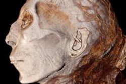
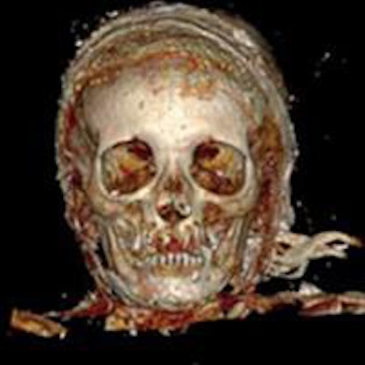
 A 3D reconstruction with volume rendering of the head, frontal view. All images courtesy of JFR.
The excavation was scheduled to coincide with groundworks undertaken for the construction of a new congress center in Rennes that would integrate the old edifice. When it was explored, the site yielded nearly a thousand graves from between the 14th and 18th centuries and different burial areas; nave and choir, adjoining chapels, cloister galleries, the chapter house, and outside the area immediately surrounding the building. Within this remarkable burial site, several lead coffins and heart-shaped urns, signs of more privileged individuals, were uncovered in the choir of the abbey.
A trapezoidal lead coffin containing a natural mummy that was particularly well preserved was thus discovered. The body could be identified from the convent archives: It was that of Louise de Quengo, Lady of Brefeillac, whose death occurred in 1656. The remains were undressed and subjected to a postmortem CT, then an autopsy. Numerous samples for spectrometric, genetic, and anatomo-pathological assessment were taken. These studies showed the absence of embalming and the intentional postmortem retrieval of the heart, which was very probably buried with her husband. The heart of her husband, Toussaint de Perrien, knight of Brefeillac, was in fact, very close to Louise's lead coffin, in a heart-shaped lead urn.
Radiological, anthropological, medicolegal, and paleo-pathological studies were carried out. Pathologies that were ante-mortem or that informed researchers about the subject's lifestyle were revealed: arterial atherosclerosis, bilateral kidney stones, and bilateral pleural adhesions as well as an intentional cranial deformity. No sign of trauma that could explain death could be visualized. Furthermore, lead diffusion within the soft tissues and the bones was depicted on CT and confirmed by spectroscopy.
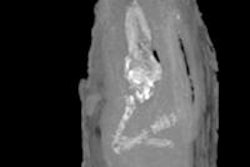
 A 3D reconstruction with volume rendering of the thorax, abdomen, and pelvis, frontal view.
This exceptional case allows a novel anthropo-archeological approach not just from traditional analysis of bones and teeth, but also the organs and soft tissue of the individual. This person, who died in 1656, generates medicolegal reflection in the same way as would a contemporary individual deceased days or weeks ago, although the material studied is 359 years old. This gives us a direct and original perspective on the relatively unknown funereal practice of removing the heart postmortem, documented not only by studying bone and cartilage, but also the surrounding organs and soft tissue.
This case illustrates the benefit and necessity of interdisciplinary collaboration in such exceptional cases. For this case, the collaboration between INRAP, CHU Toulouse (Rangueil) and the Laboratory of Molecular Anthropology and Synthesis Imaging (Laboratoire d'Anthropologie Moléculaire et Imagerie de Synthèse AMIS, UMR 5288) allowed rapid radiological exams and biological sampling and analysis.
Further collaboration with historians at Rennes University allowed us to fill in the gaps in the story of Louise de Quengo, grand noblewoman of Brittany who had been long forgotten. In a totally unexpected and unprecedented way, Louise de Quengo has taken the media world by storm. Truly, mummies from any quarter intrigue us.
Dr. Fabrice Dedouit is from the radiology department at CHU Toulouse-Rangueil, the medicolegal department, and the Laboratory of Molecular Anthropology and Synthesis Imaging (Laboratoire d'Anthropologie Moléculaire et Imagerie de Synthèse AMIS, UMR 5288), in Toulouse. Dr. Fatima-Zohra Mokrane is from the radiology department and the laboratory. Drs. Rozenn Colleter, Sylvie Duschene, Patrice Gerard, and Eric Crubezy are from the laboratory. Drs. Frédéric Savall, Daniel Rouge, and Norbert Telmon are from the lab. Dr. Hervé Rousseau is from the radiology department.
Editor's note: This is an edited translation of an article published in French on 18 October 2015 by le Quotidien des JFR (Journées Françaises de Radiologie Diagnostique et Interventionnelle), the daily newspaper of the French national congress of radiology. Translation by Frances Rylands-Monk.