Growing collaboration between researchers in Germany and Brazil is helping to propel MRI of the lung into the clinical arena and increase the level of global interest in the technique's potential.
"Focusing on the multiparametric and functional evaluation that only MRI can give, there is a lot more to come," noted Dr. Marcel Koenigkam Santos, PhD, an attending physician at the University Hospital of Ribeirao Preto, University of Sao Paulo in Brazil. "In my institution, the first projects are focusing on COPD (chronic obstructive pulmonary disease) patients and evaluation of lung cancer. Prediction of pulmonary function before surgery and early response to cancer conservative therapy at the moment is our hot topics. But we are beginning to plan research in other areas, like quantitative evaluation of interstitial diseases with MRI."
 Dr. Marcel Koenigkam Santos, PhD
Koenigkam Santos and his colleagues in Sao Paulo are performing lung MRI examinations, including evaluation of nodules and other lesions. The clinicians and surgeons have learnt about indications and applications of the method, and it is becoming part of their clinical evaluation more and more frequently, he explained.
He is overseeing several ongoing research projects involving pulmonary MRI, which could only be accomplished after doing his postdoctoral degree at Heidelberg University in Germany, thanks to a scholarship from the Brazilian government (CAPES, ministry of education). He was based at Heidelberg between February 2012 and July 2013, and during the stay, he conducted research in thoracic radiology and took part in the hospital's daily routine, practical activities, multidisciplinary meetings, etc.
"It was a great opportunity to meet a lot of people and visit places, to make new contacts and more friends," said Koenigkam Santos, who is also head of chest imaging and also coordinator of the radiology residence program.
While he was in Germany, he published nine original articles in peer-reviewed journals (six as first author), two case reports, and two review articles and gave several presentations at congresses. He received the young investigator scholarship from the Organizing Committee of the 3rd World Congress of Thoracic Imaging (WCTI) and the Magna Cum Laude at the 3rd WCTI for his presentation on noncontrast-enhanced preoperative assessment of lung perfusion in patients with non-small-cell lung cancer using Fourier decomposition MRI. He says his long-term aim is to keep developing pulmonary MRI, in order to show that it is a good method to evaluate almost all pulmonary pathologies.
Latest study on lung MRI
Koenigkam Santos' latest article was posted as an article-in-press by the European Journal of Radiology on 23 October, and was co-authored with colleagues from the Department of Diagnostic and Interventional Radiology at the University of Heidelberg.
"In this study, we propose a technique for contrast-enhanced MRI of the chest that can be applied in the clinical scenario," he explained. "It allows us to visually evaluate enhancement pattern/progression and to obtain quantitative perfusion analysis of lung lesions within the same method. A volumetric-interpolated T1-weighted GE sequence, already available in most commercial MRI systems, is applied within a strategy adapted to the pulmonary circulation."
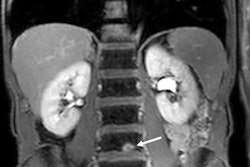
 A 63-year-old man smoker, with a < 3.0 cm indeterminate nodule in the middle lobe. CT image (lung window), T2-weighted morphological MR image, diffusion-weighted imaging (DWI) (b = 800) and one contrast-enhanced VIBE image. The morphological MR images can easily depict the nodule's shape and contours, as is demonstrated on CT. This nodule showed higher restriction signal on DWI and a prominent and fast enhancement on dynamic postcontrast images, with an early peak of enhancement > 15% and maximum enhancement > 40%, suggesting malignancy. It was a pulmonary squamous cell carcinoma. Image courtesy of Dr. Marcel Koenigkam Santos, PhD.
The aim was to propose a technique for evaluation of pulmonary lesions using contrast-enhanced MRI and to assess morphological patterns of enhancement and correlate quantitative analysis with histopathology. Volumetric-interpolated T1-weighted images were obtained during consecutive breath-holds after bolus triggered contrast injection.
Volume coverage of first three acquisitions was limited (higher temporal resolution) and the last acquisition was obtained at the 4th minute. Two radiologists individually evaluated the patterns of enhancement. Region-of-interest-based signal intensity (SI)-time curves were created to assess quantitative parameters.
Over six months, 36 patients (18 men; mean age, 62 years; age range, 40-82 years) with focal lung lesions were prospectively recruited for this study. Patients with pulmonary nodules > 1 cm and masses that had clinical indication to histopathological investigation (surgery or biopsy) were included. Patients with any therapeutic intervention prior to scanning, any contraindication to perform MRI, or to paramagnetic contrast media were excluded.
Three patients presented with more than one lesion that could be included, and therefore a total of 39 lesions were evaluated. In 27 patients, diagnosis was obtained after surgical resection, while in seven patients it was obtained by means of biopsy (endobronchial or percutaneous). In two patients, there was no pathological confirmation because indication to an interventional diagnostic procedure was re-discussed after examination, and diagnosis was assigned after follow-up exams.
All studies were performed on a 1.5-tesla MR syestem (Siemens Healthcare Aera) using a body phased-array coil (eight elements), without any external gating device or patient preparation.
A fast volumetric sequence with high spatial and contrast resolution was used to perform contrast-enhanced MRI of focal lung lesions within a strategy adapted to the pulmonary circulation, according to the authors. Quantitative analysis was highly sensitive to detect malignancy, in agreement to what has been described for other imaging methods as dynamic CT and PET.
The lack of consensus
"So far, there is no consensus on the best way to perform postcontrast images of chest MRI in a clinical scenario," they wrote, adding that it can be done in a similar fashion as it is routinely performed for abdomen/pelvis examinations, but it should be adapted to pulmonary circulation and should consider previous experience in evaluation of pulmonary lesions by imaging. A simpler and robust technique for postcontrast images can help bring MRI into daily clinical application, as an alternative or complementary tool for evaluation of pulmonary lesions.
Koenigkam Santos believes this study represents another step forward in bringing lung MRI into the clinical scenario. Since it was performed, further advances have been made, especially to make the VIBE sequence faster and more robust -- e.g., with "CAIPIRINHA" acceleration -- and he is convinced the method is going to be improved again soon.
The EJR paper is the last article he wrote based on data acquired in Heidelberg. He is still in touch with his German colleagues, however, and they are planning further collaborative projects. The plan is to strengthen the partnership between the institutions.