A research project that began as a comparative investigation into stationary and moving atomic clocks has led to the development of a new type of cardiac imaging device. Speaking at the U.K. Institute of Physics' Medical Physics Group's recent Translational Techniques meeting, Ben Varcoe, PhD, described how a collaborative effort bridged the gap between the lab and the clinic.
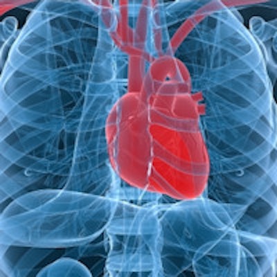
Varcoe, a quantum physicist at Leeds University in the U.K., explained how his initial atomic physics experiment led to the emergence of a device with sub-femtotesla sensitivity to magnetic fields. He exploited this capability to create a magnetocardiography (MCG) device, which records the local magnetic fields generated by a beating heart's electrical activity.
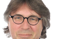
 Ben Varcoe with project manager Leanne Burgin and PhD student Shima Ghasemi Roudsari.
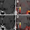
By using the MCG to map this magnetic signal at intervals over the chest, a distinctive image is created, deviations in which can indicate the presence of cardiac disease. Varcoe presented example MCG data in which images of healthy hearts were easily distinguished from images of hearts with unstable angina (which can indicate an ensuing heart attack) or non-ST-elevated myocardial infarction (NSTEMI, a potentially fatal type of heart attack), conditions that are difficult to detect with existing techniques such as ECG.
While MCG has the potential to provide complementary or superior diagnostic information to ECG, which measures the electrical activity of the heart, Varcoe notes that currently, "MCG devices are spectacularly expensive."
What's needed is a low-cost and portable system that could be used in accident and emergency departments to quickly assess patients presenting with chest pains. In the U.K., such patients are admitted to hospital for tests that can take up to three days. "If we could have an MCG device that works in an unshielded environment, we could get a diagnosis in less than three hours," Varcoe said.
With this goal, the Leeds team has developed a compact, inexpensive, portable magnetometer that -- unlike other MCG systems -- does not rely on superconducting quantum interference devices (SQUIDs), which must be operated within shielded rooms. "Our device can record images in five minutes, works through clothing, is relatively easy to use, and is now ready for the clinical environment," Varcoe said.
But the story doesn't end there. A working prototype in a university laboratory cannot simply be taken over to a nearby clinic and tested on patients. David Brettle, PhD, head of medical physics and engineering at Leeds Teaching Hospitals, explained what happened next.
Enter the medical physicist
Brettle described the life cycle of any emerging medical device. This involves initial idea generation, prototype design and development, in-house testing, large-scale production, and, finally, marketing and clinical use. The route to clinical implementation, however, can be stymied by the innovation "valley of death," a well-known phenomenon when establishing a new product or company.
This valley of death results from factors such as gaps in funding, problems in understanding medical device regulations and standards for hospital environments, or even just lack of access to colleagues with appropriate skills in other disciplines. Brettle worked with Varcoe and his team to overcome these hurdles and bring the MCG device from the lab into the clinic.
Input from the medical physics team was particularly invaluable in the areas of compliance, regulation, and patient safety. Brettle and colleagues performed assessments to ensure that the magnetometer met the technical standards for safety and effectiveness of medical electrical equipment (IEC 60601), and risk management requirements for medical devices (ISO 14971).
The medical physics team also undertook a hazard analysis. Even though the device was noncontact, Brettle deemed the proposed design -- in which the magnetometer was suspended above the patient -- as a crush risk. Other design flaws such as finger trap risks and trip risks were identified and preventative measures established.
Brettle, together with Leanne Burgin, PhD, from the University Innovation and Knowledge Centre, also negotiated the issues surrounding CE Marking and Medicines and Healthcare products Regulatory Association (MHRA) registration of medical devices, establishing that neither was required to begin noncommercial, research-based investigations of the device.
Clinical testing
Thanks to the efforts of the medical physics team and clinical colleagues, the Leeds MCG is now undergoing initial clinical testing under Dr. Mark Kearney, professor of cardiology at the Leeds General Infirmary. The first study was designed to assess the magnetometer's technical performance and determine whether it can differentiate between images of normal and abnormal hearts.
The researchers are examining three groups: healthy volunteers; patients with NSTEMI or ischemic heart disease (neither of which can normally be detected using an ECG); and those with ST-elevated myocardial infarction (STEMI, a severe form of heart attack). Typical MCG maps for each group were easily distinguished from each other. Varcoe pointed out that in addition to classifying images as healthy or unhealthy, the system appears to be able to differentiate between different cardiac conditions.
In terms of diagnostic ability, the device is demonstrating exceptional potential for specificity and sensitivity in differentiating healthy volunteers from post-MI patients using the magnetic field map.
The team is also investigating intra- and interoperator reliability and reproducibility in a patient over seven days. "So far, the device is working exactly as we want it to," Varcoe said. "From a fundamental physics beginning, we have developed a new medical device."
© IOP Publishing Limited. Republished with permission from medicalphysicsweb, a community website covering fundamental research and emerging technologies in medical imaging and radiation therapy.