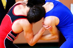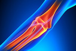
It's official: MRI was the overwhelming winner when it came to elbow investigations on competitors at the 2012 Olympic Games. Also, most elbow injuries occurred in power and combat sports such as judo, boxing, wrestling, and weightlifting, according to one of the first peer-reviewed, scientific articles about the games.
To mark the first anniversary of the London Olympics, the first wave of articles about the imaging aspects are starting to emerge. Hot on the heels of an overview article that was published online on 23 July by the British Journal of Sports Medicine, a detailed scientific analysis about the incidence and evaluation of elbow injuries at the games will appear in the September edition of the American Journal of Roentgenology.
"Elbow injuries in Olympic sports have not been reported previously," noted lead author Dr. Sarath Bethapudi, from the department of musculoskeletal radiology at Leeds Teaching Hospitals National Health Service (NHS) Trust in the U.K. "Most of the injuries resulted from valgus strain with hyperextension of the elbow and usually presented as injuries to the medial joint supporting structures. Combinations of medial and lateral ligaments were seen in combat and power sports with high-energy acute trauma. Such injuries also resulted in tears to secondary stabilizers of the medial joint, including the common flexor tendons and medial muscular compartments" (AJR, September 2013, Vol. 201:3, pp. 535-539).
 A 30-year-old male judo athlete with valgus stress and hyperextension injury of elbow. Top: Coronal proton density-weighted fat-saturated MRI shows full-thickness tear (white arrow) of proximal humeral attachment of anterior bundle of ulnar collateral ligament (UCL) and edema (black arrow) within surrounding muscle fibers. Below: Axial proton density-weighted fat-saturated MRI shows UCL posterior bundle disruption (long white arrow), with fluid surrounding ulnar nerve (short white arrow). However, ulnar nerve was contiguous on sequential images, with no transection. High-grade tear of flexor carpi ulnaris (black arrow) also was seen. Bottom: Axial proton density-weighted fat-saturated MRI shows high-grade tear of flexor digitorum superficialis (long arrow) and fluid surrounding median nerve (short arrow). All images reprinted with permission from the American Journal of Roentgenology.

A total of 1,711 radiological investigations were performed at the main Olympic Village polyclinic, of which nearly 50% were MRI exams. Of these investigations, 36 were performed on 30 elbows in 28 athletes, including 26 MRI exams, nine ultrasound scans, and one CT scan. Two athletes underwent MRI of both elbows. Two examinations were of poor quality because of metal artifacts and patient movement. Of the MRI exams, 22 were abnormal, showing at least one pathologic abnormality. Eight of the nine ultrasound scans were abnormal and the lone CT exam confirmed an avulsion fracture, as suspected on MRI.

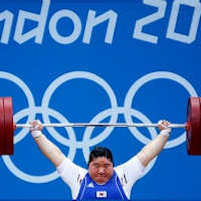
 A 20-year-old male weightlifter with flare-up of chronic elbow pain. Coronal proton density-weighted fat-saturated MRI (left) and coronal proton density-weighted nonfat-saturated MRI (right) show anterior bundle UCL midsubstance tear (arrows). Absence of significant edema within disrupted fibers and adjacent soft tissue suggests that tear is chronic.
Of the 28 athletes, 15 were male and 13 female, and the mean age was 25 years (standard deviation 5.2 years), according to Bethapudi, whose co-authors came from the International Olympic Committee in Lausanne, Switzerland, and the department of orthopedic surgery at Oslo University.
Judo and weightlifting were the two largest sporting groups scanned, he added. Of the 30 elbows scanned in the athletes' category, 28 were for sports-related injuries, whereas two of the scans were for acute nonsporting trauma.

 A 22-year-old female boxer who sustained direct blow to elbow during bout. Left: Radiograph of elbow shows avulsion fracture (arrow) of lateral epicondyle. Right: Coronal proton density-weighted fat-saturated MRI shows radial collateral ligament avulsion (short solid arrow). Note edema of lateral epicondyle at site of avulsion (open arrow), complete tear of proximal UCL, and superficial common flexor tendon (long solid arrow).
 The same patient. Left: Coronal proton density-weighted fat-saturated MRI shows high-grade tear of lateral UCL (LUCL) humeral attachment (short arrow). Distal attachment of LUCL at supinator crest of ulna (arrowheads) is intact. Complete tear of UCL humeral attachment and common flexor attachment (long arrow) is seen. Right: Axial proton density-weighted fat-saturated MRI shows avulsion fracture of lateral epicondyle (long arrow). Extensive high-grade muscle tear of anterior and medial muscular compartments (short arrow) also is shown.
Of the 28 elbows scanned for acute sports injuries, 15 had high-grade ligament injuries, and 12 of these ligament injuries occurred in contact sports and weightlifting. The remaining three injuries were seen in throwing athletes, two of whom were javelin throwers and one of whom was a volleyball player.
"Tears of the common flexor and extensor tendons occurred in combination with ligamentous injuries. This occurs because common flexors and extensors act as secondary stabilizers of the elbow joint and are injured when the primary stabilizers fail. Once again, these injuries occurred primarily in combat sports, weightlifting, and overhead-throwing athletes," stated Bethapudi, who was responsible for data collection and analysis at the 2012 Olympics and has received funding from GE Healthcare, a sponsor at the games.


 A 27-year-old male handball athlete with acute and chronic elbow pain. Coronal (left), sagittal (middle), and axial (right) proton density-weighted fat-saturated MRI examinations show osteochondral injury of capitellum (arrows). In contrast to posterior capitellar impaction resulting from acute dislocation, radial head shows no fracture or edema. Findings are in keeping with isolated chronic osteochondral injury. It is difficult to ascertain whether osteochondral injury is acute or chronic according to imaging alone.
Significant ligament and tendon injuries to the elbow can occur often in nonthrowing athletes, and most of the elbow injuries seen in these athletes were isolated high-grade ulnar collateral ligament (UCL) injuries, although combinations of medial and lateral ligament injuries can occur, the authors concluded. Ulnar attachment tears were the next most common injuries, followed by midsubstance tears of the UCL. This trend differs from the existing literature, which suggests that midsubstance tears are the most common type of UCL injuries, they wrote.
Interestingly, they think there's still scope for studying the biomechanics of elbow injuries in nonthrowing athletes at international sporting events.
Of the 1,711 investigations at the 2012 games, 48.8% were MRI exams, 20.2% were diagnostic ultrasound scans, 23.6% were plain radiographs, 2.9% were CT scans, and interventional procedures accounted for 4.3%, according to the same authors' article in the British Journal of Sports Medicine. Nearly 75% of imaging was performed on athletes, while less than 5% of the services were utilized by the workforce. Demand on radiology services peaked during week two of the games.