DUBAI - Most PET/MR systems cost between $5 million and $7 million U.S. (3.7 million and 5.2 million euros), whereas a new generation PET/CT machine with 128 detectors costs between $1.5 million and $2 million U.S. (1.1 million and 1.5 million euros). The key question is whether PET/MR is worth the extra dollars, a global expert on hybrid imaging told delegates at Arab Health, which began today.
 The Arab Health exhibition and congress opened its doors for the 38th time on Monday in Dubai. The main focus this year is on diabetes and complementary medicine, but the 13th Middle East Medical Imaging and Diagnostics Conference is being held during the event.
"I certainly do see it as a value player and differentiator. Over time, price is going to fall," said Dr. Shetal Shah, co-director of PET services at the Imaging Institute of the Cleveland Clinic in Cleveland, Ohio, U.S. "If you look from the patient's perspective, this is really the right thing to do for evaluation. It's something that's been coming and has already arrived. It'll be up to us as early adopters to prove its clinical benefit and superiority over PET/CT."
PET/MRI combines the exquisite detail and soft-tissue contrast resolution of MRI with the superior sensitivity of PET for detection and characterization of disease, and is becoming a valuable tool for focused applications that can reduce cumulative ionizing radiation, he said. Now early adopters are working on improving clinical workflow and throughput, optimizing protocols, working on reimbursement, and developing the necessary technical and clinical expertise.
"It's kind of like coming out with the very first iPhones, when people didn't rush in lines to get in," Shah said. "Is there a cost-benefit analysis? Is it more cost-effective, and so forth? That's what's probably going to happen over the next two or three years. A lot of publications are going to come out and show PET/MR's clinical usefulness as well as cost-effectiveness."
Turf issues, and specifically who will interpret PET/MR images, also must be addressed. No formal guidelines exist in this area, but with training in PET, anybody who has clinical expertise in MRI should be able to interpret these images, he thinks, and the best way to avoid disputes is for radiology and nuclear medicine to work together and make a joint effort.
 You may have learnt MR, but PET/MR is a different thing, according to Dr. Muhammad Chaudhry, medical director at the Tawam Molecular Imaging Center in Al Ain, United Arab Emirates.
Four years ago, the Cleveland Clinic created an in-house hybrid imaging team of 15 physicians with expertise in both modalities. They ensured the requirements of the Society of Nuclear Medicine (SNM) and American College of Radiology (ACR) were met, and these physicians comment on each other's interpretations and diagnoses and help each other regularly.
"With the growth of techniques and utilization, the more people you have working together -- one with expertise in PET, one with expertise in MR -- you'll see a clear benefit," noted Dr. Muhammad Chaudhry, medical director at the Tawam Molecular Imaging Center in Al Ain, United Arab Emirates, who spoke at the same session on Monday. "You may have learnt MR, but PET/MR is a different thing. Not everybody is looking at the same things, so you have to have all kinds of expertise available."
As for the choice of PET/MRI system, the overriding practical consideration tends to be space availability, Shah noted. If only a small amount is available, then the best option is the simultaneous acquired system from Siemens Healthcare, but if the true time-of-flight detector capability of PET is required, a large footprint is available and a purchaser needs to save about $1 million U.S. (740,000 euros), then the Philips Healthcare TF product is probably the solution, he said.
Significantly, Philips is also working on a simultaneous machine, so in another year or two, simultaneous acquisition will win out, according to Shah.
 About 80,000 attendees are expected at this week's Arab Health. More than 40,000 delegates have preregistered, which is around 10% more than last year, according to the organizers.
A basic whole-body PET/MR takes between 35 and 40 minutes, and is essentially a replacement product for, say, a pediatric patient with lymphomas who must undergo repeated scans four or five times a year, he explained. For a more advanced imaging case involving one-stop-shop for a patient with colorectal carcinoma, for example, and where full staging is required, an exam will take 1.2 hours. At the Cleveland Clinic, two hours is allowed for such a patient.
In the presequel space following abdominoperineal resection (APR) for colorectal cancer, 80 patients at the hospital have been studied post-APR, and the key point is to find the carcinoembryonic antigen (CEA) levels. If the CEA is elevated, then a PET/CT scan must be done first to confirm the location and guide the biopsy, Shah said.
"If the patient's CEA level is negative and there is soft-tissue thickening in the pelvis, then most of the time it's fibrosis. Go to your surgeon and say, 'Listen, this is most likely fibrosis, let's wait three months and get a follow-up,' " he recommended.
In the oncologic setting, whole-body diffusion-weighted MRI is making progress, but there are still more questions than answers. Fusing a whole-body diffusion MR image with a PET scan is one area of research that will need to be looked at over the next five years, Shah concluded.
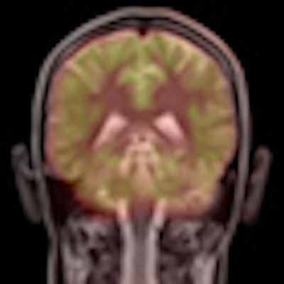
Note: The PET/MR image on the home page was obtained with the Biograph mMR PET/MR system. Image courtesy of Siemens.