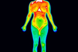
Functional 3D infrared imaging (3DIRI) displays promise as a breast cancer risk assessment tool, reportedly offering high sensitivity and reasonable specificity, according to a proof-of-concept study published online in European Radiology.
Israeli researchers developed a prototype high-resolution 3DIRI device, using an imaging protocol with temporal resolution coupled with multiparametric computer analysis for providing risk assessment of breast cancer. Their hypothesis was that the 3DIRI device would detect tumors' metabolic signatures and help clinicians assess a woman's breast cancer risk.
Early detection and prevention strategies for breast cancer depend on the ability to accurately identify individuals with significantly increased breast cancer risk, wrote lead author Dr. Tamar Sella of Hadassah Hebrew University Medical Center in Jerusalem and colleagues. But current risk assessment models are statistical and rely on clinical features such as genetic susceptibility, family history, or mammography breast density. Functional imaging such as 3DIRI could offer information on the metabolic nature of the breast tissue and better distinguish tumors from healthy tissue (Eur Radiol, December 6, 2012).
"The technology presented in this study attempts to provide a dynamic assessment analyzing the chances that a woman harbors a breast cancer at a specific time, based on objective metabolic signatures," Sella and colleagues noted. "If assessed as high-risk, such a woman would need immediate further workup to detect and localize the cancer."
3DIRI is appealing because it is noninvasive, does not expose women to radiation, and potentially causes less patient discomfort as data are collected, the authors stated. They emphasized that the technology is not infrared thermography, which is based on finding asymmetric thermal hot spots that are thought to represent malignancy.
The device prototype consisted of dual optical heads set up at 60° angles between each other and at a distance of 68 cm from the patient (who was seated upright throughout).
- The optical heads are stationary and cover an area of 44° x 24.5°.
- Together, the two heads can produce a 3D infrared map of the breast at a maximum imaging frequency of 9 Hz.
- The infrared imager operates between the 7.5-µm and 13.5-µm infrared band.
- The optical heads are calibrated every day for a uniform signal baseline.
Between September 2009 and July 2011, 1,827 women were examined with 3DIRI at one of six breast imaging centers. According to particular criteria, Sella's team identified and included 434 of these 1,827 women and divided them into two subgroups. The first group included 256 women who had a negative routine screening mammogram (category 1 based on BI-RADS); these women were imaged with 3DIRI immediately before the mammogram and served as the control group. The second subgroup included 178 women who had an image-guided breast biopsy with a result of either invasive breast carcinoma or ductal carcinoma in situ, and were the cancer cohort of the study.
The researchers randomly divided the 434 participants in the study within each subgroup into two further subsets: a training and calibration set that included the first 204 healthy women and 112 women with breast cancer, followed by a blinded test set that included 52 healthy women and 66 with breast cancer.
"The initial training and calibration phase was designed to determine the optimal threshold for each descriptor and to determine the contribution of each descriptor in the overall diagnostic performance for risk analysis of breast cancer," the authors wrote. "During the calibration process, various thresholds were tested for each algorithm separately, defining the optimal threshold to be used."
An initial 10-minute period of temperature stabilization preceded the imaging process, during which the patient sat in the dedicated imaging room with room temperature set at 68° F (20° C). Following the temperature equilibration phase, continuous infrared imaging was taken for five minutes. The imaging session total time was about 15 minutes.
Screening mammography exams were taken with Selenia Dimensions units (Hologic) and a Senographe 2000D device (GE Healthcare).
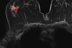
The study found that the sensitivity and specificity of functional infrared imaging in providing the correct risk for the presence of breast cancer were 90.9% and 72.5%, respectively. Forty-two of the 60 cancers, or 70%, in women correctly classified by the system as suspicious were smaller than 20 mm in size, according to the researchers.
"The specificity of 3DIRI for correctly assessing a woman as high-risk among our select population was 72.5%," they wrote. "This evaluation is limited by the fact that the absolute standard of reference in the current study for healthy control cases was mammography, which in itself is an imperfect technique for breast cancer depiction, especially in women with dense breasts."
The team acknowledged that their study had some limitations, such as its design as a two-armed blinded study that included only healthy woman or those diagnosed with cancer and excluded all other lesions, which could impede the evaluation of the technology's true performance and introduce a possible inherent selection bias. And as this study is the first clinical performance test for 3DIRI, the group has not yet investigated the effect of benign lesions of the breast on false-positive rates and of precancerous lesions on false-negative rates.
Yet this initial data suggest the technology merits further investigation, according to Sella's team, since developing an accurate, simple, noninvasive, radiation-free technology that estimates the chances that a woman has breast cancer has the potential to help in both breast cancer screening and workup of questionable lesions.
"Theoretically, future clinical applications could include prescreening prior to mammography, short-term follow-up of high-risk women, evaluation of young women below the age of 40, or assessment in women with equivocal findings to help determine whether further workup should be applied," the group wrote. "Since the technology is completely automated, its potential benefit is further enhanced, making it an easy and accessible tool for physicians."