Low-dose unenhanced CT scans may underestimate the volume of smaller lung nodules compared with standard-dose contrast CT studies, creating potential problems when tracking nodules over time, wrote researchers in the October issue of the American Journal of Roentgenology.
The findings indicate that clinicians performing follow-up on suspicious nodules should keep in mind that nodules may seem to be different sizes based on the type of CT protocol used. Nodule volumes were from 13% to 15% lower on low-dose unenhanced CT exams compared to standard-dose contrast-enhanced CT exams for nodule volumes 200 mm3 and smaller (p < 0.001), but no significant differences were seen in larger nodules.
"This effect is likely due to contrast administration rather than other imaging parameters, which should be taken into account in the follow-up of lung nodules because growth can remain undetected or doubling time underestimated," wrote Dr. Pim de Jong and colleagues from University Medical Center Utrecht in the Netherlands (AJR, October 2012, Vol. 199:4, pp. 777-780).
The number of lung nodules of unknown etiology is increasing with greater use of MDCT, and the volume of incidental nodules brings daunting challenges for physicians charged with distinguishing malignant from benign lesions, the authors wrote. Absent a few obvious clues to benignity such as full calcification, fat density, small size, or perifissural location, clinicians are left to rely on slow volume doubling times of 600 days or less to define potentially malignant solid nodules.
The Fleischner guidelines for lung nodule management rely on average nodule diameter on axial CT to measure size, and they recommend low-dose CT, which has shown results comparable to standard-dose scanning. To help manage all of the nodules and boost reporting accuracy, volumetric measurement of nodules is gaining favor. Unfortunately, the presence of contrast may make nodules appear larger than those scanned without contrast, according to the authors.
Not only in other studies but "in our own practice, measured nodule volume seemed to be dependent on the use of IV contrast material in combination with the applied radiation dose," they wrote.
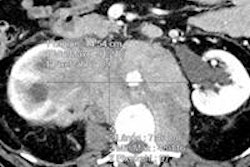
The current study aimed to compare volumetric nodule size between standard-dose contrast-enhanced scans and low-dose unenhanced scans. The research team scanned 20 patients with known pulmonary metastases three times in a single day: two unenhanced low-dose scans and a standard-dose contrast-enhanced scan of the chest.
Chest CT scans were performed on either 16- or 64-detector-row systems (Mx8000 IDT, Brilliance 16P, or Brilliance 64, Philips Healthcare) at collimation of 16 x 0.75 mm or 64 x 0.625 mm, respectively. The group reconstructed axial images for analysis at 1-mm (16-detector-row) or 0.7-mm increments (64-detector-row) for all studies. Low-dose unenhanced images were acquired at 120 kVp and 30 mAs, and 60 to 85 mL of IV contrast was injected prior to contrast-enhanced scans at 120 to 140 kVp and 75 to 200 mAs, depending on body weight.
The group used a portable workstation (IntelliSpace, Philips) and the vendor's automated software to measure nodules smaller than 1,000 m3, and for significant differences, Wilcoxon's signed rank tests were used to analyze nodules ≤ 200 mm3 and > 200 mm3.
In all, 101 nodules were analyzed in 15 subjects. The measured volumes of nodules ≤ 200 mm3 (69 nodules) were systematically lower on both low-dose unenhanced CT exam protocols compared to standard-dose contrast-enhanced CT (differences, 13.7% and 15.5%; p < 0.0001). However, nodule volume did not significantly differ between low-dose CT (median difference, 1.0%; p = 0.10) or between the protocols for larger nodules > 200 mm3 (p > 0.30).
Especially for smaller nodules ≤ 200 mm3, representing the majority of incidental detections, variation between protocols can be large and the growth rate underestimated, the study team wrote, concluding that low-dose unenhanced CT underestimates volume by about 15% compared to standard-dose contrast-enhanced CT.
And that 15% can have important clinical implications, because it's often large enough to spell the difference between a nodule that is worked up or one that is left alone due to apparent lack of growth in the interval between scans, they wrote.
"Let us assume a malignant nodule of 100 mm3 (approximately 5.8 mm) with a volume doubling time of 300 days," de Jong and colleagues wrote. "This nodule is detected on a baseline standard-dose contrast-enhanced CT study. When this nodule is followed up after 100 days with standard-dose contrast-enhanced CT, the newly measured volume would be 126 mm3, a significant increase of 26% and a malignant growth rate (doubling time, 300 days). However, when this nodule is followed up with low-dose unenhanced CT, the new volume would be only 107 mm3, a nonsignificant increase of 7% and a benign growth rate with a doubling time of 969 days."
As for the reason behind the volume differences, the authors hypothesized that it is an effect of contrast enhancement, as differences have been shown in previous studies between contrast and noncontrast CT. It appears that the increased nodule density caused by contrast uptake alters the edge detection of the software, leading to larger volume measurements, they said.
A difference of 15% in nodule size has important implications "for the introduction of volumetry in the clinical setting because malignant growth may remain undetected when low-dose unenhanced CT is used for follow-up of detected nodules in clinical practice when the prior CT was contrast enhanced," the group concluded.