VIENNA - High-pitch chest CT is a robust technique that provides high-quality thoracic images of babies in motion -- quickly, without sedation, and at low radiation doses -- according to a study presented at the European Congress of Radiology (ECR).
Still, high-pitch dual-source CT also comes with more overranging, so the resulting greater anatomic coverage edged those high-pitch radiation doses higher than they would have been using a comparable conventional CT system, and were in fact equivalent to it, the results showed.
Using CT in young children, "sedation or intubation is required to improve image quality, particularly in patients who are small and cannot comply with instructions," said Dr. Michael Lell, from University Clinic in Erlangen, Germany. "So our aim was to transfer the [high-pitch CT] protocol that we know from cardiac scanning to the pediatric population."
The study of 60 young patients (31 male, 30 female, mean age 15 months) examined with thoracic CT included 30 scanned using a high-pitch dual-source CT protocol (Definition Flash, Siemens Healthcare) and 30 scanned using conventional low-dose helical CT (Sensation 10 or 64, Siemens Healthcare). There was free breathing during the scans and no sedation.
- Conventional CT images were acquired using pitch of 1.3, 10 or 64 x 0.6-mm collimation, 80 kV (100 kV for > 10 kg body weight), 50 effective mAs, and 0.75-mm reconstruction interval.
- Dual-source CT images were acquired in either single- or dual-source mode using pitch of 1.3 or 3.0, 128 x 0.6-mm collimation, 80 kV, (100 kV for > 10 kg body weight), and 0.75-mm reconstruction interval.
Using a standard soft-tissue edge-enhancing kernel, image quality was assessed using a four-point scale (0 = no artifacts, 3 = severe artifacts), and comparative dose expressed in CTDIvol and organ doses.
Radiation dose (mSv)
|
The average radiation dose was 1.9 ± 0.6 mSv for conventional CT and 1.9 ± 0.6 mSv for high-pitch-mode dual-source CT. The mean scan time was 0.49 seconds in high-pitch mode and from 3.5 to 7.6 seconds using conventional CT.
Doses did not differ significantly between the scanning techniques, and, in particular, images acquired using high-pitch DSCT did not yield lower radiation doses "due to the broader range of overscanning -- the area that we irradiate but do not reconstruct," Lell said.
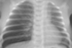
However, image quality was superior using high-pitch mode owing to a significant reduction in motion artifacts compared to 10- and 64-detector-row conventional CT, he said.
Image quality comparison
|
Among patients scanned with conventional CT, artifacts were seen at the level of the diaphragm (100%), the borders of the heart (100%), the ribs (67%), and spine (20%). In the high-pitch mode artifacts were seen in only six patients in the lung parenchyma, next to the diaphragm or the heart (p < 0.001), the authors wrote in their abstract.
"We can obtain excellent image quality without any sedation at very low radiation exposure with high-pitch mode, and this turns out to help a lot in terms of workflow because we do not have to call in the patients before in order to get them in contact with the anesthesiologist," Lell concluded. "There's no comorbidity because of this intubation or sedation, and there is no follow-up period so this imaging can be performed on an outpatient basis."
Due to these advantages, the study team uses high-pitch mode on all of its very young patients, he said.
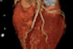
In response to a question from the audience, Lell said the proximal coronary arteries could be seen using the technique, but a full evaluation of the coronary arteries isn't possible due to high heart rates that typically range from 100 to 120 bpm.