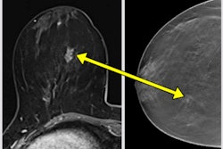
Contrast-enhanced mammography (CEM) confirms the effectiveness of ultrasound-guided cryoablation and could serve as a reliable annual follow-up exam, findings presented on 26 November at RSNA 2023 suggest.
In her presentation, Dr. Federica di Naro from Azienda Ospedaliero-Universitaria Careggi in Florence, Italy, discussed her team’s results, which showed that combining CEM and re-evaluation with biopsy in selected breast cancer histology proves the efficacy of ultrasound-guided cryoablation and CEM’s reliability.
“CEM is a valuable tool to define the effectiveness of cryoablation,” di Naro said.
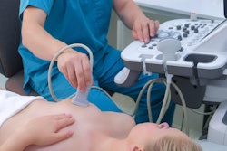
Cryoablation is a minimally invasive treatment option that sees targeted tissues killed by extreme cold, using ultrasound guidance. Di Nara noted that this is a useful alternative for elderly women not eligible for surgery. She added that CEM is also a useful imaging modality for elderly women who may struggle to stay still or maintain specific positions needed for MRI.
Di Nara and colleagues wanted to find out whether ultrasound-guided cryoablation could be effective in treating low-risk early-stage breast cancer, combining CEM and re-evaluation with biopsy.
For the study, the team included data from cryoablation data from 28 biopsy-proven malignant lesions in 28 patients with an average age of 73.4 years. The patients were held inoperable by a multidisciplinary group, under hormone therapy. The team reported that all lesions were ultrasound-visible invasive ductal carcinoma (< 30 mm), low to intermediate grade, HR-positive, and HER2-negative.
 Federica di Naro, MD, from Azienda Ospedaliero-Universitaria Careggi in Italy discusses her team's results at RSNA, showing that contrast-enhanced mammography (CEM) confirms the effectiveness of cryoablation and can serve as a valuable follow-up exam tool.Amerigo Allegretto
The patients underwent CEM staging to confirm the unifocality of the lesions and were subdivided according to molecular subtype, Ki-67 percentage, and 1-cm dimensional cutoff. The researchers analyzed cryoablation outcomes via the size reduction rate with ultrasound at one, three, and six months, and with CEM and biopsy at 12 months. Histology results served as the gold standard.
The team found that 28 lesions had an average reduction rate of 20.1% at one-month follow-up. This increased to 66.4% at three months and 94% at six months. The team also reported that hyperechoic procedural scarrings were visible without any recognizable ultrasonic trace of the index lesion in 13 lesions after three months, and in 25 lesions after six months. Of the total 28 lesions, 14 were Luminal-A and 14 were Luminal B Her-2 negative.
| Ultrasonic characteristics of lesions undergoing cryoablation over time | |||
|---|---|---|---|
| Reduction rate | 1 month | 3 months | 6 months |
| Luminal-A | 21.6% | 70.2% | 96.8% |
| Luminal B | 19.8% | 60.7% | 93.4% |
| Breast cancer > 1 cm | 19.7% | 63.4% | 93% |
| Breast cancer < 1 cm | 23% | 84.4% | 100% |
| Ki-67 > 20% | 18.8% | 62.8% | 96.5% |
| Ki-67 < 20% | 24.6% | 65.3% | 93.4% |
For the CEM part of the study, the team found no detectacle enhancement in 26 of the total lesions. It reported that the two lesions with enhancement presented mainly as mass. In these cases, biopsy confirmed residual traces of B5 lesions only in the core zone of the scarring site, corresponding to CEM results. In all the negative CEM cases, biopsy reported only benign lesions.
Di Naro said that these results point to ultrasound-guided cryoablation being a safe, feasible, and effective breast cancer treatment method. She added that the results demonstrate CEM’s necessity for follow-up exams.