PET/MRI shows promise in diagnosing fevers or inflammation of unknown origin and may have advantages over PET/CT, according to a study published on 3 January in the European Journal of Radiology.
In a study that included 104 patients, PET/MRI provided high sensitivity and specificity for determining underlying causes, especially in patients with elevated C-reactive protein, a blood biomarker of inflammation, according to lead author Dr. Tomas Rohan of University Hospital Brno in the Czech Republic.
“To make the indications more specific to and improve the overall diagnostic performance, F-18 FDG-PET/MRI should be indicated in FUO and IOU especially in patients with elevated CRP,” the authors noted.
Fever of unknown origin (FUO) is defined as a temperature higher than 38.3 °C lasting more than three weeks and of which the cause has not been determined during more than three visits to the doctor or three days in the hospital. Inflammation of unknown origin (IUO) is defined as prolonged and perplexing inflammation with temperatures below 38.3 °C.
Both FUO and IUO can be caused by a wide range of pathological processes, from malignancies, infections, and granulomatous and inflammatory diseases, to miscellaneous causes. Moreover, if the etiology persists undiagnosed for more than six to 12 months, the likelihood that a specific diagnosis will ever be made decreases, the authors explained.
PET/MRI is a relatively new hybrid method being gradually integrated into clinical practice, with no experience using F-18 FDG-PET/MRI to diagnose FUO and IUO reported in the literature, they noted.
To address this knowledge gap, the group retrospectively analyzed F-18 FDG-PET/MRI scans of 104 patients with FUO or IUO who underwent imaging at their hospital between July 2016 and January 2021. They evaluated the sensitivity, specificity, and predictive values of the PET/MRI findings in relation to the final diagnosis.
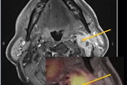
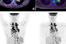
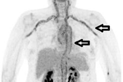
 In an example image from the study, F-18 FDG-PET/MRI fusion image (A) and PET 3D image (D) show a moderately increased FDG uptake in muscles around hip joints. T2 IDEAL WATER image (B) shows increased T2 signal; LAVA WATER (C) image shows mild enhancement in these muscles. Findings were suspicious of myositis, which was histopathologically confirmed. Image courtesy of the European Journal of Radiology.
Furthermore, the researchers assessed clinical (fever, arthralgia, weight loss, night sweats, and age) and laboratory (C-reactive protein, leukocytes) parameters and compared these with the true positivity rate of PET/MRI.
Based on agreements between two radiologists and two nuclear medicine physicians, FDG-PET/MRI achieved a sensitivity of 96 %, specificity of 82 %, and positive and negative predictive values of 92% and 90 %. The cause of FOU or IUO was determined in 71 patients (68.3 %) and included such causes as polymyalgia rheumatica, adult-onset Still´s disease, vasculitis, pericarditis, and giant cell arteritis. In 33 (31.7%) patients, the etiology of IUO/FUO remained unknown.
Also, the most significant parameter in relation to subsequent PET/MRI finding was increased level of C-reactive protein, which was present in 96% of true positive PET/MRI cases, the researchers added.
Ultimately, while PET/CT imaging has proven highly valuable in helping identify causes for FUO and IUO, the method involves higher radiation exposure to patients than PET/MRI, and thus may not be as suitable a choice for older, more sensitive patients, the researchers wrote.
“F-18 FDG-PET/MRI is a suitable alternative for the investigation of fever and inflammation of unclear etiology. This imaging technique has especially high sensitivity, negative predictive value and low radiation exposure,” the group concluded.