High-resolution SPECT is playing an invaluable role in small-animal imaging, a tool for the development of new therapeutic strategies for human medicine. The extremely high-resolution, artifact-free images produced by this modality are making it feasible to track the response of pharmaceuticals in animal models of cancer and diabetes.
Freek Beekman, PhD, a professor of radiation, detection, and medical imaging at TNW Delft University of Technology in the Netherlands, discussed the use of SPECT technology for small-animal imaging in a video interview with medicalphysicsweb. In the video, he described recent achievements in improving SPECT resolution from a clinical value of 1 cm to 0.35 mm in a living mouse.
 
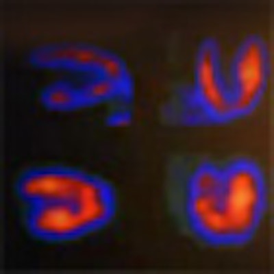
Freek Beekman, PhD, describes how high-resolution SPECT systems play an invaluable role in small-animal imaging. Video courtesy of medicalphysicsweb.
|
Beekman also discussed the work of MILabs, a company that he founded to commercialize preclinical molecular imaging. MILabs has developed a range of SPECT systems, including the VECTor system that can perform simultaneous SPECT and PET studies. This combined PET/SPECT device exploits clustered multipinhole collimator technology to deliver submillimeter resolution.
Looking to the future, Beekman said that it should be possible to translate the SPECT technology from the preclinical arena into human imaging. He predicted that such a system could, for example, perform cardiac imaging with two to three times higher resolution than currently possible, with equivalent scanning time and dose. Or, it should be possible to achieve high image quality using far less radioactive material, he noted.
© IOP Publishing Limited. Republished with permission from medicalphysicsweb, a community website covering fundamental research and emerging technologies in medical imaging and radiation therapy.