Individuals with long COVID-19 show decreased gray-matter volume on brain MR imaging compared to healthy controls, Hungarian scientists have reported in the Journal of Magnetic Resonance Imaging.
The results provide new information about the effects of long COVID, according to Gábor Perlaki, PhD, and colleagues from the University of Pécs.
"The most characteristic symptoms of long COVID persisting beyond six to 12 months may suggest an involvement of the central nervous system: physical and mental fatigue, migrating/multiplex pain complaints, persistent olfactory disturbance, sleep disturbance, dyspnea, cognitive and concentration impairment ('brain fog')," they wrote in an article published on 22 August.
COVID-19 manifests primarily as a respiratory infection, but up to 40% of patients who contract the illness develop late and prolonged symptoms, including neurological ones, but previous studies have offered contradictory findings regarding these neurological symptoms, the researchers stated in article published on 22 August in the Journal of Magnetic Resonance Imaging.
"After the acute illness has resolved, some patients may retain some of their symptoms for a longer time, or even develop new symptoms," they wrote. "The causes of these symptoms are unclear. Some hypotheses suggest that cardio-pulmonary damage, persistent viral infection, postinfectious immune response, postinfectious transient autonomic dysfunction, psychological, and psychosocial factors may be involved in the development of the symptoms ... [which] can cause neurological symptoms even without direct central nervous system involvement."
The group sought to investigate chronic effects of COVID-19 on gray matter in a cohort of 38 young patients (average age, 26) without comorbidities who experienced mild COVID and had no medical complaints at the time of assessment; this cohort's results were compared with 37 controls who had not contracted COVID-19. All study participants underwent an MRI exam at least 60 days after their first positive test for COVID-19, received neuropsychological testing, and completed a questionnaire.
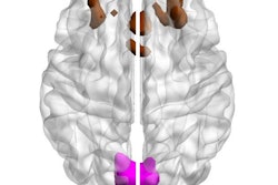
The researchers found that the COVID cohort had significantly lower bilateral mean cortical thickness, lower subcortical gray matter, and lower right olfactory bulb volume compared with the healthy controls.
| Brain measures between healthy controls and those with COVID-19 on MR imaging | ||
| Measure | Healthy controls | COVID cohort |
| Lower bilateral mean cortical thickness | 2.6 mm | 2.5 mm |
| Lower subcortical gray matter | 60,470 mm3 | 57,881 mm3 |
| Lower right olfactory bulb volume | 61 mm3 | 52.3 mm3 |
The investigators also found that in patients with moderate to severe olfactory function loss, cortical thickness was significantly lower compared to patients without it, at 2.6 mm versus 2.5 mm. They also reported reduced cortical thickness in the right lateral orbitofrontal cortex in the COVID group compared with the healthy control group (2.6 mm vs. 2.5 mm).
The takeaway? Long COVID sufferers may not show symptoms, the team noted.
"Even without any subjective or objective neurological complaints at the time of the MR scan, subjects in the COVID group showed gray-matter alterations in cortical thickness and subcortical gray-matter volume," the authors concluded.
The complete study can be found here.