Ultrasound-guided vacuum-assisted excision is not a reliable method for removing small breast cancers, according to research from Finland published on 16 August in the European Journal of Radiology.
A team led by Michaela Björnström from the University of Helsinki found that none of the small invasive breast cancers identified in their study were completely removed with vacuum-assisted excision under ultrasound guidance. However, they also found that after visible lesions have been excised, there are still small foci of cancer and ductal carcinoma in situ (DCIS) left, meaning "larger amount of tissue than visible on ultrasound needs to be removed for complete lesion excision."
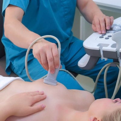
With advancements in imaging technology such as screening mammography, cancer detection comes earlier when tumors are smaller. While it's currently challenging to determine which of these small tumors will become malignant, vacuum-assisted excision under local anesthesia is a method that safely removes tumors.
Previous research suggests that this method is successful in removing nonmalignant lesions. However, Björnström and colleagues pointed out a lack of data on whether vacuum-assisted excision is reliable for removing small breast cancers. The team wanted to add to the literature, investigating whether it's possible to completely remove small breast cancer tumors with ultrasound-guided vacuum-assisted excision in patients.
The researchers included 10 women with an average age of 64 years who had histologically confirmed breast cancer 10 mm or smaller on ultrasound and mammography. The median size of the tumors on mammography was 8.5 mm and 6.5 mm on ultrasound. The researchers also reported that of the total lesions, three were round, two were oval, and five were irregularly shaped. Also, the median breast thickness at the site of the lesion was 25 mm.
The Björnström and colleagues reported that none of the tumors were completely removed in the first excision specimen. They wrote that this means that there was invasive cancer or DCIS in the shaved margins and/or the surgical specimen. The team also found that in five cases, the surgical specimen was free of invasive cancer and DCIS.
However, the study authors wrote that while their study size was small, they could not proceed with the study since none of the 10 first tumors treated with vacuum-assisted excision could be successfully removed.
They added that "most" of the lesions in the study were located in a fatty part of the breast. They reasoned that breast tumors are heavier than surrounding fat tissue, making it harder for the vacuum to suck out the tumor.
"Furthermore, slowly growing tumors can consist of tight fibrotic tissue and may therefore be more difficult to remove by vacuum-assisted excision," the authors wrote.
The full study can be found here.