Photon-counting CT enhances image quality for coronary CT angiography exams while also reducing contrast use by up to 40%, Swiss researchers have stated. The technology's higher contrast-to-noise ratio properties are of particular note, they said.
The findings are good news for patients, since reductions in contrast media may help them avoid complications from coronary CT angiography (CCTA) exams, noted a team led by Dr. Giulia Cundari of the University of Zurich in Switzerland. The group's findings were published on 31 July in Academic Radiology.
"Increased image quality of photon-counting CT (PCCT) can be used for considerable contrast media volume reduction while still maintaining a diagnostic image quality of CCTA," the team wrote.
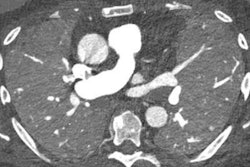
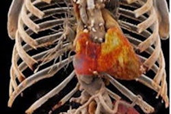
CCTA is the go-to imaging modality for evaluating coronary artery disease. But the exam carries risk of contrast media-induced nephropathy -- making it important to use the least amount of contrast possible without compromising image quality. And reducing contrast volume also has other benefits, including mitigation of the environmental effects of the breakdown of iodine and potential contrast media shortages due to supply chain disruptions.
PCCT appears to offer a solution, reducing the volume of contrast media without negative effect on the quality of the images. To explore PCCT's performance in this regard, Cundari and colleagues conducted a study that included data from 100 patients who underwent a CCTA exam with dual-source PCCT.
The participants were divided into three groups: group 1 (35 patients), which was used to set the optimal virtual monoenergetic image (VMI) energy level (determined to be 40 keV); group 2 (also 35 patients), which underwent the exam at a 20% reduced contrast volume; and group 3 (30 patients), which underwent the exam at another 20% lower contrast dose.
There were no significant differences in age, body mass index, body weight, blood pressure, heart rate, or estimated glomerular filtration rate between the groups.
The group's contrast media ranges depended on the flow rate (mL per second) of administration of the contrast.
- Group 1 contrast media range: 72 mL to 85.2 mL
- Group 2 contrast media range: 58 mL and 68 mL
- Group 3 contrast media range: 43.2 mL and 51.6 mL
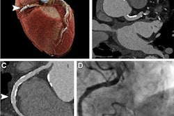
The investigators tracked the following image quality measures: vascular attenuation, image noise, and contrast-to-noise ratio. Two readers blinded to the VMI energy level and contrast protocol rated images for overall quality, subjective image contrast, and subjective noise using a five-point scale.
Interreader agreement was good to excellent for each group (kappa, 0.62 to 0.92). The best overall image quality was obtained at 45 keV, the investigators noted.
| Mean contrast-to-noise ratio at 45 keV (with higher values equal to more noise) | |
| Group | Mean contrast-to-noise ratio |
| 1 | 32 |
| 2 | 25.8 |
| 3 | 20.5 |
At 45 keV, overall image quality scores were high, at 4 out of 5 on the rating scale for group 1, 5 on the rating scale for group 2, and 4 on the rating scale for group 3, the team reported.
"45 keV was selected as the ideal energy level for CCTA, providing the optimal trade-off between objective and subjective image quality," the authors noted.
The research results highlight how PCCT can significantly lower contrast requirements on CCTA, according to Cundari and colleagues.
"Our study demonstrates the possibility to reduce the volume of contrast media for CCTA by 40% while maintaining a diagnostic image quality ... [by] exploiting the lower noise and higher contrast-to-noise ratio properties of low-energy-level VMI reconstructions with [PCCT]," the team wrote.
The complete study can be found here.