Researchers have overcome an inability of ultrasound to map capillaries and other vessels in the brain by injecting micrometer-sized bubbles into the bloodstream. Although the technique has been demonstrated only on a model brain so far, it promises to be a simple, inexpensive way to provide a complete mapping of a real brain's vascular networks (Medical Physics, November 2013, Vol. 40:11, pp. 110701-110707).
The mapping of blood vessels in the brain is known to be an effective tool in studying diseases such as Alzheimer's. Current technology, however, makes vascular mapping difficult. CT scans have a resolution of about 400 micrometers (µm), which rules out all but the largest blood vessels. MRI using higher field strengths of 3 tesla to 7 tesla can reveal more vessels, but this is a costly process that is rarely available. More common, clinical MRI, on the other hand, has a resolution of 300 µm.
Ultrasound imaging ought to be a higher-resolution alternative. But ultrasound cannot penetrate the skull at those higher frequencies that afford greater resolution, and as a result, the imaging technique has been able to map only major blood vessels behind thinner sections of the skull. One option has been to seek help from the field of ultrasound therapy, which employs a hemispherical array of transducers to improve the signal. "This works to a point, but the low frequency used still gives relatively poor resolution," said Medical Physicist Kullervo Hynynen, PhD, at the Sunnybrook Research Institute in Toronto, Ontario.
With his colleague Meaghan O'Reilly, Hynynen has found that ultrasound imaging can be improved with superresolution. This technique has been used in optics to increase resolution beyond the so-called diffraction limit, by relying on tiny bright spots of radiation positioned within a region being imaged. In Hynynen and O'Reilly's ultrasound version of superresolution imaging, they use micrometer-sized bubbles of gas, which reveal their position by strongly scattering the ultrasound back to the receiver array.
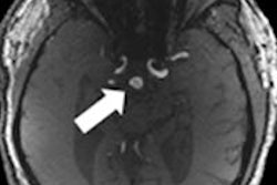
The researchers tested their technique on a model brain that consisted of an ex vivo skull containing a blood vessel made of 255-µm-wide plastic tubing, and found they could still map the vessels with ultrasound. In principle, the researchers say, the resolution could be as great as the bubble size -- that is, 1 µm to 3 µm -- which would allow the mapping of capillaries.
In a real brain, Hynynen said, the bubbles would be cleared by the lungs and liver in a matter of minutes. He and O'Reilly are currently performing animal testing. "We estimate that this could be tested in patients within a few years," he said. "The method can be used for other organ imaging, [but] brain imaging is actually more difficult than other organs."