Dual-source CT (DSCT) scanners provide accurate measurements of iodine concentration across a wide range of cardiac CT protocols with the use of dual-energy CT (DECT) myocardial perfusion imaging protocols, a Dutch-led research team wrote in European Radiology.
In a study that compared DECT-derived iodine concentrations to true concentration in phantoms representing different patient sizes, the results showed excellent correlation between the two measurements, the study team wrote.
The mean measurement errors were approximately 3% for first-generation DSCT scanners, and 2.9% for second-generation machines, reported Dr. James D. Koonce, and Dr. Rozemarijn Vliegenthart et al from the University of Groeningen in Holland, and Dr. U. Joseph Schoepf, Dr. Felix Meinel, and colleagues, from the Medical University of South Carolina in Charleston (Eur Radiol, 20 October 2013).
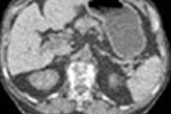
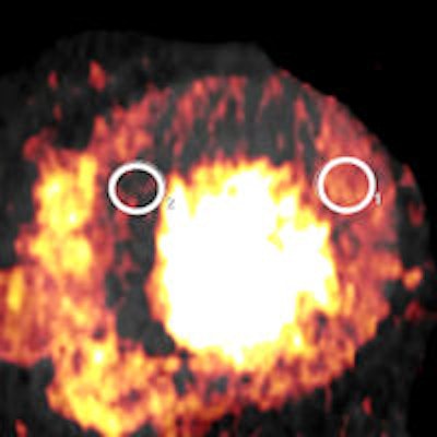
 A 45-year-old man with stenosis of the left anterior descending artery (LAD). The image in the top row was obtained at rest and in the bottom row during pharmacological stress. DECT showed normal iodine concentrations during rest, but at stress the iodine concentration within the antero-septal myocardium is decreased with a quantitative iodine concentration measurement of 0.6 mg/mL compared with 3.4-3.8 mg/mL in healthy myocardium at rest and stress. Clinical images not from phantom study courtesy of Dr. Felix Meinel.
"We found that dual-energy CT overall allows for accurate measurements of iodine concentrations across cardiac CT protocols, strengthening the case for dual-energy-CT-derived blood volume estimates as a surrogate of myocardial blood supply," Meinel wrote in an email to AuntMinnieEurope.com.
To date, studies on cardiac dual-energy CT focused on the subjective visual evaluation of DECT iodine maps for the presence of myocardial iodine distribution defects as signs of ischemia or infarction, Meinel explained. This study attempts to quantify those observations to determine the accuracy of iodine measurements.
"If myocardial iodine concentration can be accurately measured based on dual-energy-CT, this may allow for the definition of specific iodine concentration cut-offs for normal and abnormal perfusion and for an estimation of myocardial flow reserve in patients with coronary artery disease undergoing rest and stress dual-energy CT," Meinel wrote.
The accuracy of myocardial perfusion estimates depends on how well dual-energy cardiac CT quantifies iodine concentrations, a topic that has not been investigated to date, according to the authors.
This study aimed to assess the accuracy of dual-source CT scanners for quantifying iodine concentration in a thoracic phantom model using various cardiac dual-energy CT acquisition protocols across a range of simulated patient sizes, Meinel wrote.
The scans were performed using both first- and second-generation DECT systems (Somatom Definition and Somatom Definition Flash, Siemens Healthcare) in dual-energy mode.
The researchers equipped an anthropomorphic phantom with tubular inserts containing known iodine concentrations from 20 mL in the cardiac chamber, and fat rings to simulate different patient sizes. Dedicated software was used to measure iodine concentrations at DECT and the results were compared to true concentrations. Linear regression models to identify predictors of measurement accuracy.
Multiple protocols, two scanner models
First-generation DSCT scans were obtained in spiral and acquisition modes, using 330 ms gantry rotation time, pitch 0.2 in spiral modes, detector collimation of 2 x 32 x 0.6 mm, and slice acquisition 2 x 64 x 0.6 mm with z-flying focal spot technique. One tube was operated at 150 mAs and 140 kVp, the second at 165 mAs and 80 or 100 kVp, and no tube current modulation was used.
In the second-generation DSCT scanner, all tube outputs were set to 100 kVp; flying Z-spot technique permitted operation in 64- and 128- slice modes, and a tin filter was used in conjunction with 140 kVp in order to improve spectral separation. DECT acquisition variables allowed six different protocol combinations, the study team wrote.
The iodine concentrations derived from DECT were measured using dedicated software and compared with true concentrations placed in the phantoms, and linear regression models were used to identify predictors of measurement accuracy.
The results showed excellent correlation between measured and true iodine concentrations in 72 datasets across both CT systems and multiple protocols ( r = 0.994 - 0.997, p < 0.0001). The mean measurement errors between real and measured CT values were 3.0 ± 7.0 % for first-generation scanners and -2.9 ± 3.8 % for second-generation dual-source CT.
The group also reported a significant (p = 0.002) interaction between true iodine concentration and phantom size, and measurement accuracy decreased with increasing phantom size. The mean error was -0.8 ± 3.9% without the fat ring simulating larger patients, 2.0 ± 4.9 % when a five-cm fat ring was used, and 8.3% when a 10-cm fat ring was used. And overall accuracy of iodine measurements was better for 100 kVp than for 80 kVp, especially at lower iodine concentrations, the study team wrote.
High accuracy overall
"We demonstrated that the overall accuracy of iodine measurements using first- and second-generation DSCT in DECT image acquisition mode is very high among all protocols that are typically applied in cardiac imaging," the group wrote, adding that the small differences in accuracy between protocols can be explained by technical considerations. "At 100 kVp, more radiation dose is available than at 80 kVp, if the same mAs is used as in our study... This leads to less image noise in particular for larger patient sizes and therefore to lower variability at 100 kVp."
The reduced accuracy for first-generation scanner protocols using 80 kVp, especially for larger phantom sizes is consequential, they wrote. As a result, for larger patients and iodine concentrations less than 10 mg/mL, the low energy tube spectrum should be set to 80 rather than 100 kVp.
Full reconstructions obtain twice the data as half reconstructions, and are less sensitive to scattering and other artifacts. However, temporal resolution with full reconstruction, and as a result the "disadvantages may outweigh a 2% accuracy gain" found clinically, Koonce et al wrote.
A bonus of the technique is good overall accuracy for the quantification of iodine in cardiac DSCT protocols, which the authors called "an important step towards studying the diagnostic and prognostic value of objective quantitative myocardial iodine measurements in a clinical setting."
As for limitations, the authors cautioned that scanning phantoms is inherently different from scanning patients, and variations caused by breathing, myocardial contraction, and other movements do not impact phantom results. Also, the results do not include the newer rapid kV switching technique that was not available when the study began.
"DECT provides accurate measurements of iodine concentration using cardiac acquisition protocols and different simulated patient sizes," they concluded. "The second-generation DSCT system showed the most stable measurements across a wide range of iodine concentrations and simulated patient sizes."